HUMB1000 Lecture Notes - Lecture 26: Endoneurium, Motor Neuron, Epineurium

Document Summary
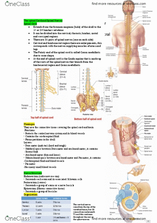
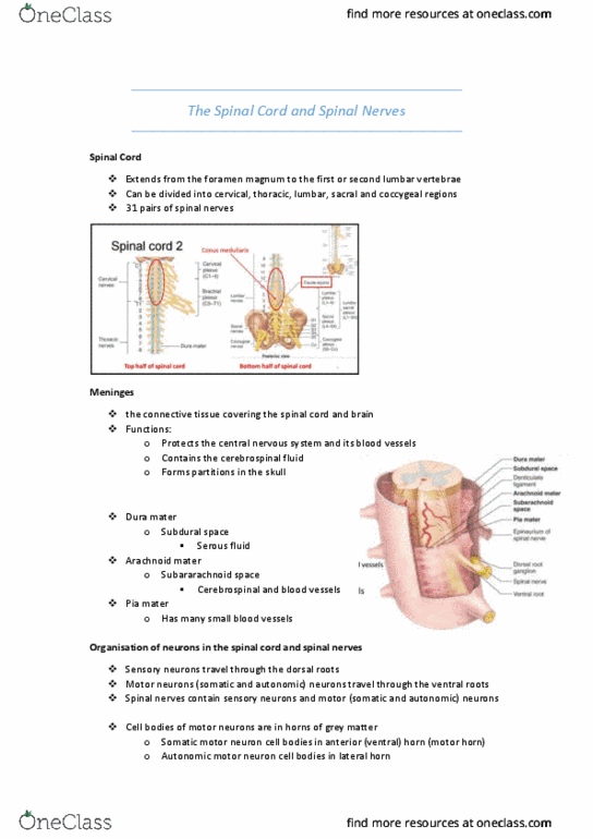
L1 - the spinal cord & spinal nerves. Spinal cord: extends from the foramen magnum to the first or second lumbar vertebrae, can be divided into cervical, thoracic, lumbar, sacral, and coccygeal regions, 31 pairs of spinal nerves, diameter changes from top to bottom. Has "enlargements" in the cervical region and lumbosacral region. Meninges: he connective tissue covering the spinal cord and brain, functions: Protects the central nervous system and its blood vessels. Forms partitions in the skull: dura mater. Looks like a cobweb: pia mater. Compendum 9 - how does it all work page 98. Somatic motor neuron cell bodies in anterior (ventral) horn (motor horn) Autonomic motor neuron cell bodies in lateral horn. Compendum 9 - how does it all work page 99. Surrounds each axon and its associated schwann cells: perineurium. Surrounds a group of axons or a nerve fascicle: epineurium - outermost. Organization of spinal nerves: cervical, thoracic, lumbar, sacral, coccygeal.