MMED2931 Lecture Notes - Lecture 6: Pulmonary Circulation, Depolarization, Medulla Oblongata

4 Jun 2018
School
Department
Course
Professor

Cardiovascular System Lectures Outline:
1. Heart structure and function
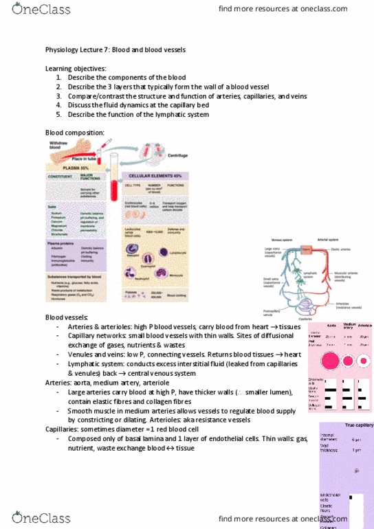
2. Blood and blood vessels
3. Control of blood flow and blood pressure
Cardiovascular System Lecture 1: Heart structure and Function
Learning objectives:
- Describe the anatomy of the heart
- Name the heart valves. Describe their location, function, & mechanism of operation
- Understand the components of the conduction system of the heart
- Describe the functional unit of contraction in the cardiac muscle cell and explain how
a contraction comes about
- Describe pressure changes in the heart during the cardiac cycle
- Define cardiac output and explain how it is regulated
Location of the heart:
NOTE: apex – the lowest point. Base – upper part
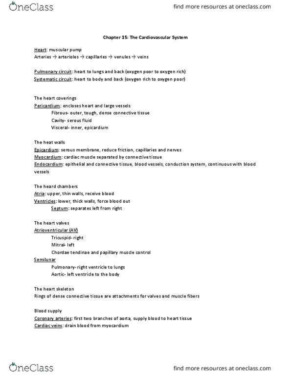
Covering and layers of the heart wall:
- Endocardium: contains endothelial tissue with small blood
vessels and bundles of smooth muscle
- Myocardium: contains cardiac muscle cells
- Epicardium: made of squamous epithelial cells overlying
connective tissue
- Pericardium: layer of connective tissue around the heart. Fixes
the heart to the mediastinum, gives protection against
infection, provides lubrication for heart
o Fibrous pericardium: outer tough layer comprising fibrous tissue
o Serous pericardium: thin, smooth. 2 layers: parietal layer (lines inside of the
fibrous pericardium), visceral layer (adheres to the surface of the heart)
o PERICARDIAL SPACE: separates parietal/visceral layers of the pericardium
o Contains 10-20mL of clear pericardial fluid that cushions the heart
Chambers of the heart: Blood vessels of the heart:
find more resources at oneclass.com
find more resources at oneclass.com

The valves in the heart:
- Semilunar valves:
o Prevent backflow into the ventricles when
ventricles relax
o Aortic semilunar valve
o Pulmonary semilunar valve
- When ventricular P > BP in aorta & pulmonary trunk, semilunar valves open
Cardiovascular system overview: heart is 2 side-by-side pumps
- Right side: pump for the pulmonary circuit
o Vessels that carry blood to/from the lungs
o Lower resistance compared with systemic circuit
- Left side: pump for the systemic circuit:
o Vessels that carry blood to/from all body tissues
Heart anatomy:
- Contraction F generated by myocardium (muscle of heart)
- Atrial walls are thin
- Left ventricle wall thicker – greater P generated by left
ventricle compared to right ventricle
Microscopic anatomy of cardiac muscle:
- Cardiac muscle cells: striated, short, fat, branched, interconnected
- Branching cardiac muscle cells held together by connective tissue (collagen, elastin
fibres)
- T-tubules: wide but less numerous. SR: simpler than in skeletal muscle
- Numerous large mitochondria (25-35% of cell volume)
-
- Cardiac muscle cells connected as a network by intercalated discs where
cell membranes are closely opposed
- Desmosomes prevent cells from separating during contraction
- Gap junctions (ensure heart contracts as unit) allow ions to pass;
electrically couple adjacent cell.
find more resources at oneclass.com
find more resources at oneclass.com