PSYC10003 Lecture Notes - Lecture 9: Muscle Spindle, Stretch Reflex, Quadriceps Femoris Muscle

14 Jun 2018
School
Department
Course
Professor

MBB1 – Lecture 9
The sensorimotor system
Muscle innervation by motor neurons of spinal cord
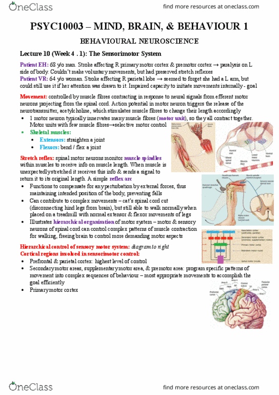
• Movements are controlled by muscles that contract in response to neural signals
from efferent motor neurons projecting from the spinal cord
o Motor neurons exit the spinal cord via the ventral root and terminate on
individual muscle fibres
• An action potential in a motor neuron triggers the release of acetylcholine, which
stimulates muscle fibres to change their length accordingly
• A single motor neuron usually innervates many muscle fibres – when neuron fires, all
the muscle fibres contract together
o Group of fibres innervated by a single neuron is called a motor unit
▪ Motor units with the fewest muscle fibres (e.g. face and hands) allow
the greatest degree of selective motor control
• Many skeletal muscles fall into one of two categories: extensors and flexors
o Fleors at to ed or fle a joit, hile etesors at to straighte i
o These categories often act antagonistically, as is the case for biceps and
triceps
Stretch reflex
• Spinal motor neurons receive input from a variety of sources, including sensory
receptors within the muscles themselves
• Activity of skeletal muscles is monitored by receptors called muscle spindles
o These provide info to the CNS regarding muscle length
• When a muscle is stretched unexpectedly (e.g. hammer taps under patella), the
muscle spindles convey info back to the spinal cord via the dorsal roots
• Axons of spindle afferent neurons synapse directly with the motor neurons, which
increase their activity in order to return the muscle to its original length
o Results in a brisk contraction of the quad muscles, causing the lower leg to
extend – circuit forms a simple reflex arc
• Functional significance of reflex is subtle – role is to compensate for any
perturbation by external forces and thus maintain the intended position of the body
o E.g. bumped while holding hot coffee, stretch reflex compensates
automatically and prevents you spilling it
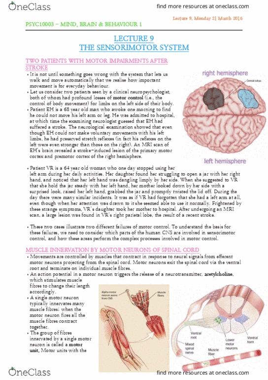
• Patients with profound loss of motor control on left side due to damage to right
hemisphere showed strong patellar tendon reflexes, indicating motor and sensory
neurons of the spinal cord remained intact
Preserved walking following spinal cord resection
• Motor neurons in spinal cord are capable of triggering quite complex movements of
various groups, without any controlling signals from the brain
• Been illustrated in experiments with cats, where the spinal cord is surgically
sectioned at a point just above where nerves serving hind legs are located
o Disconnects lower motor neurons for the hind legs from the brain
o Despite this, the cats can still walk normally on a treadmill, showing normal
extensor and flexor movements of the hind legs
find more resources at oneclass.com
find more resources at oneclass.com

• Demonstrates hierarchical organisation of the motor system – motor and sensory
neurons within the spinal cord are able to control all the complex patterns of muscle
contraction required for walking, without instructions from the brain
• Leaves brain free to control more demanding aspects of motor control
o E.g. determining when to initiate actions, which effectors to use etc.
Descending control from the brain
• The tendency to want to drop a hot dish comes from excitatory synapses of motor
neurons in the spinal cord, but this excitation can be counteracted by inhibitory
input from the PMC
• Axons that descend from the PMC via the spinal cord form inhibitory synapses with
lower motor neurons
o Similarly, excitatory inputs from the brain can trigger action potentials in
lower motor neurons and initiate movements
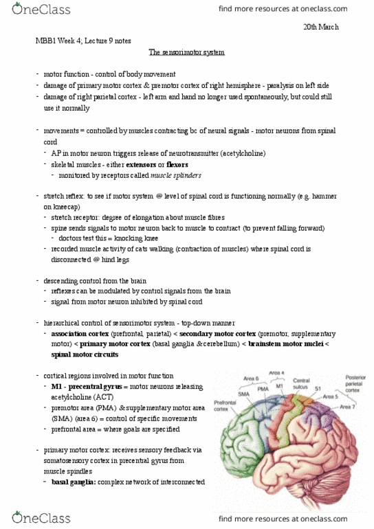
Hierarchical control in the sensorimotor system
• Association areas (prefrontal cortex and parietal cortex) specify general goals rather
than specific plans of action
o Association cortex is not routinely involved in details, leaving the highest
levels of control free to perform the most complex functions
• Areas of the secondary motor cortex (premotor and supplementary motor areas)
are involved in programming specific patterns of movement
• PMC in each hemisphere is the point of departure from which sensorimotor signals
from the brain are conveyed to the brainstem and spinal cord
• This organisation involves both top-down and bottom-up communication
o If a problem arises with one of the areas, this is conveyed back up the chain
to the higher levels, whose responsibility is to resolve any problems
o Just as sensory feedback from the muscles and tendons is monitored by the
CNS in case adjustments are required
find more resources at oneclass.com
find more resources at oneclass.com