NESC 2470 Lecture Notes - Lecture 4: Lateral Geniculate Nucleus, Amacrine Cell, Ganglion

30 Dec 2017
School
Department
Course
Professor
Document Summary
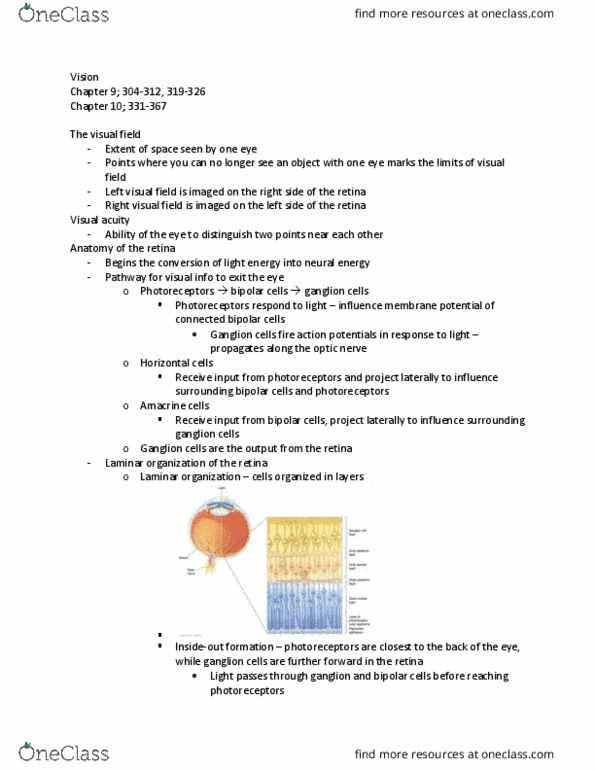
In a lamina formation: key features are the cell arrangement and where they are relative to each other. Binocular: two images, we have to merge these two images. Conscious visual pathway: retina: light enters here, signaling to the lateral geniculate nucleus in the thalamus for processing, goes towards the cortex called area 17/v1/striate cortex, expansion to other cortical regions (occipital, temporal, parietal lobes) Important for achromatic or colorless vision: cones, for phototopic vision, high acuity, combines of densities and the types of pigments within these receptors that are important for what spectrums we can detect. Fovea: where you have the most cones, the center of the retina, focal point. Important for acuity of our vision: little pit is so the light doesn"t have to pass through the cells, provides a way to get a better/clearer signal without passing though ganglion and inner/outer nuclei layer. Bind spot: no photoreceptors, where axons are leaving the eye.