BIOL 300 Lecture Notes - Lecture 8: Lac Repressor, Dna-Binding Domain, Deoxyribonuclease I

23 Nov 2017
School
Department
Course
Professor
Document Summary
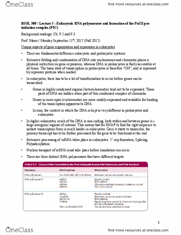
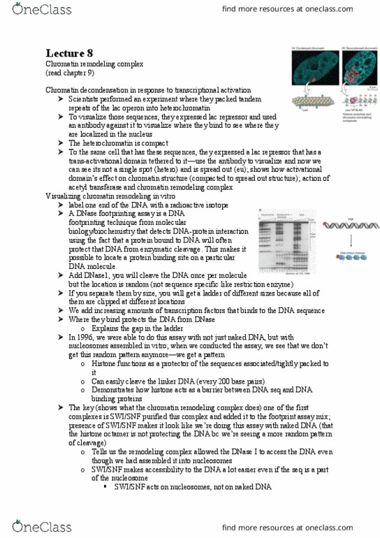
Background readings : chapter 9 lodish, and supplemental reading on mycourses (a) they specifically targeted heterochromatin regions, where chromatin is usually highly compact and in tight structure. To visualize the sequences that they introduced (which is the. Laci operon of e. coli), they used an antibody against the lac repressor (in red). The heterochromatin shows in a localized region. (b) then, to the same cell, they expressed lac repressor that was fused with an activation domain from virus. Using the same antibody, they tried to visualize where the sequences localize in the nucleus. This time, the sequences aren"t in one region, but rather in more lose conformation in the nucleus. The chromatin is not condensed anymore because of the presence of proteins binding to the activation domains. Visualizing chromatin remodeling in vitro: labeling one end of the dna with fluorescence probe. By adding dnase i in diluted solution such that it cleaves dna only in two fragments.