ANAT 261 Lecture 14: Lecture 14 - Resp

18 Feb 2019
School
Department
Course
Professor
Document Summary
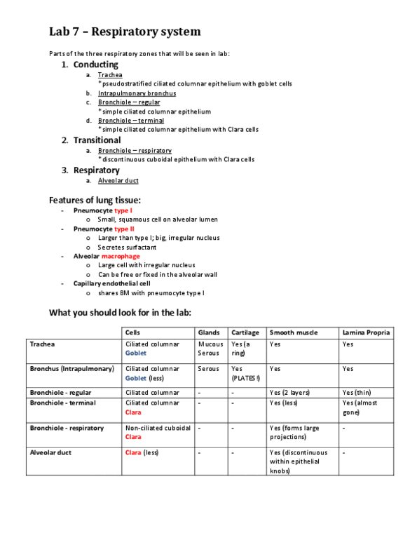
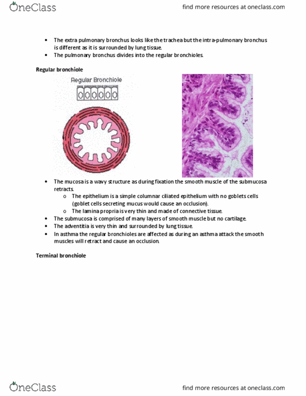
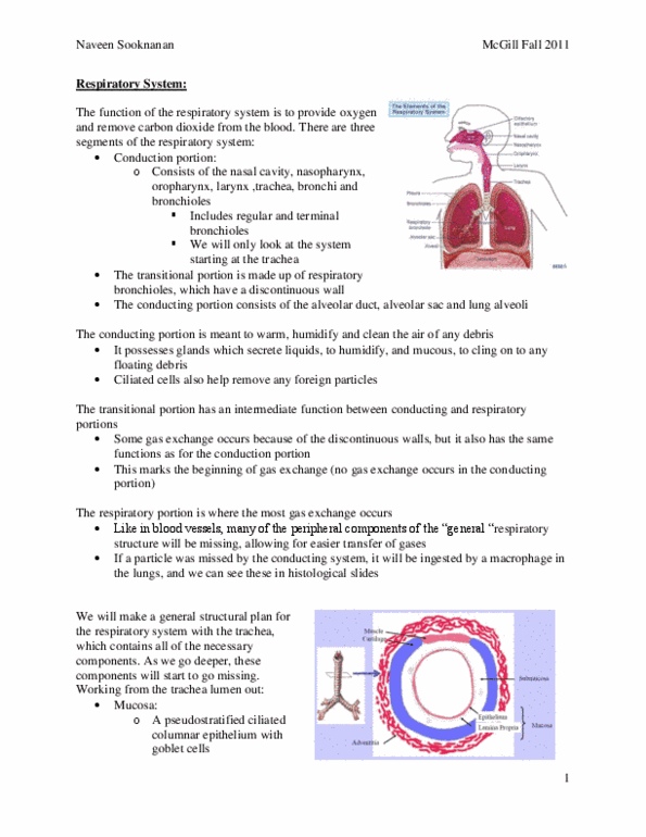
Intrapulmonary bronchus: large structure, only a portion of a cross-section (portion of a wall) can be seen in the field of vision of light microscopy. October 19th, 2017 towards the lungs (in the pulmonary bronchi you don"t have anymore: you don"t want mucous inside the smaller structures, pieces of hyaline cartilage, structure that will allow you to classify the pulmonary bronchus: Any time you see this wavy structure with a piece of cartilage you will classify the structure as the pulmonary bronchus. Note: as you move distally (towards the lungs) in the respiratory system, structures get smaller in diameter and also become more branched. If you look around, you will see a lot of lung tissue surrounding the bronchiole: has no hyaline cartilage and has no goblet cells, therefore it must be a bronchiole (can"t be mistaken for a bronchus) General plan of the terminal bronchiole (layers: mucosa layer, epithelial layer: mix of columnar ciliated cells and non-ciliated columnar.