ANAT 321 Lecture Notes - Lecture 17: Lateral Geniculate Nucleus, Optic Chiasm, Parvocellular Cell

17 Oct 2016
School
Department
Course
Professor
Document Summary
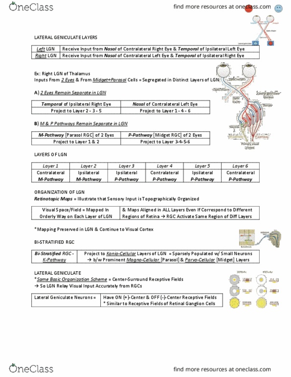
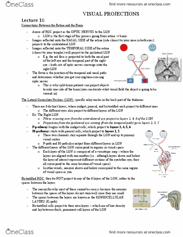
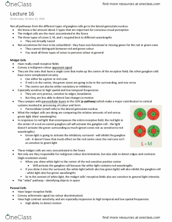
Colour vision: colour vision is due to the combination of separate (parallel) visual pathways corresponding to the three different types of cones/ganglion cells, midget, bistratified, parasol ganglion cells, the p- m- and k-pathways, respectively. Part ii: from the retina to the cortex. The axons of retinal ganglion cells project as the optic nerves to the lateral geniculate nuclei. The fibers partially cross at the optic chiasm and continue as the optic tracts: the splitting of the fibers are functionally organized. The nasal fibers cross, the temporal fibres do not: the left side of the visual field projects to the contralateral (right) side of the lgn, and vice versa. Midget and parasol ganglion cells project to six distinct layers in the lateral geniculate nucleus (lgn) The lgn is in the thalamus (caudal and ventral) It has a strikingly layered structure: cells in these layers are packed very densely.