EAST 501 Lecture 7: 7- M. Nagano

21 Jun 2018
School
Department
Course
Professor

7- M. Nagano
11- Male reproduction- spermatogenesis
1- Anatomy of the tesis
●What is the difference in gametogenesis (production of sperm and egg) between male and
female?
○Quantity: males produce lots of sperm, while females produce one egg at a time.
○Duration: for males the production is continuous (almost life-long), and for females, it is
periodic/cyclic (and is limited → menopause).
○Men: 150 million sperm per day vs 1 egg per 28 days in women.
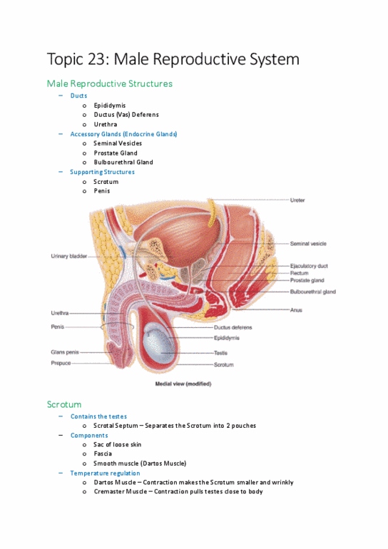
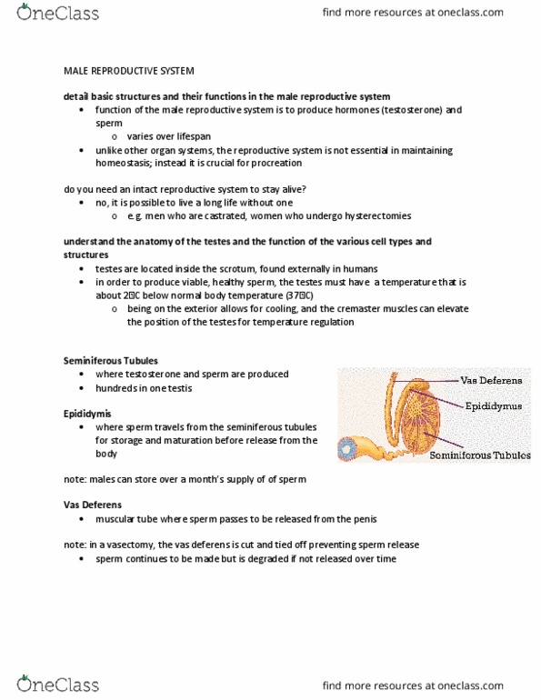
●Anatomy of the testis
○Many seminiferous tubules in the testis → epididymis → vas deferens → ejaculation.
●Morphology of testis, seminiferous tubules, and seminiferous epithelium
○Within the tubule, can find sperm at different development stages.
■Sperm cell: haploid.
■To make those haploid cells, they have to undergo meiosis. First, spermatocytes
have 4N DNA. One spermatocyte then divides into four haploid cells via 2
meiotic divisions.
○Spermatogonia at the top of the basal membrane → proliferate, and eventually commit to
undergo meiosis (spermatocytes). Then, they then become spermatids → at the bottom of
the tubule.
○Sertoli cells are the somatic cells that directly support spermatogenesis.
●Anatomy of seminiferous epithelium and interstitial tissues in the testis
○Testes have two different anatomical components: first the tubules, and then outside of
the tubules.
■Inside the tubules, find germ cells and Sertoli cells.
■Outside of the tubules, find Leydig cells and interstitial cells (blood and
lymphatic vessels). Leydig cells are the cells that produce testosterone.
○Cartoon: see the structure more clearly.
●The testis has two major functions
○Exocrine: secretion of cell components to outside of the body (sperm).
■Production of male gametes.
■Germ cells = spermatogonia, spermatocytes, spermatids, spermatozoa.
○Endocrine: inside the body, through the bloodstream → production of hormones.
■Sertoli cells: inside the seminiferous tubules. Direct regulator of germ cells.
Express LRH. Really determine sex during embryogenesis → SRY drives sexual
differentiation into the male.
●// Granulosa cells.
■Leydig cells: outside of the seminiferous tubules in interstitial tissue. Express
LRH. Support spermatogenesis indirectly via hormone production. + induction of
secondary sex characteristics.
●// Theca cells.
●Summary
○See slide.
2- Development of germ cells- spermatogenesis
1

7- M. Nagano
●Cytoplasmic bridge: clonal progression of spermatogenesis
○Spermatogonia are diploid cells and reside at the bottom of the seminiferous epithelium.
○Divide to expand the mother population. Then they enter meiosis and eventually become
diploid. While they divide, their cytokinesis is incomplete → morphological signature of
commitment to differentiation.
■Once they take his morphology, they will go all the way to becoming
spermatozoa.
○One cell can produce about 1000 cells, and then released all at once from the epithelium.
●What is the difference in gametogenesis between male and female?
○A man continuously produces ~1000 sperm at every heartbeat for almost his entire life.
●Different stage of spermatogenesis can be found along the seminiferous tubules
○Very long structure. In histological sections, depending on where you cut, you always
see different cell combinations.
■If you cut at a certain point: will see cells at different stages.
■Means that in these two meters of seminiferous tubules, the spermatozoa are
constantly released to be ejaculated in order to fertilize as many eggs as possible.
●Cycle of spermatogenesis is species-specific
○The different stages take different amounts of time depending on species.
■For men: takes about two months for completion of spermatogenesis.
■Rat: 48 days.
●Summary
○See slide.
3- Endocrine communication that regulate spermatogenesis
●Hypothalamus- pituitary- testis axis
○Reproductive endocrinology takes places in this major axis. Spermatogenesis is regulated
by protein and steroid hormones in this HPT axis.
●Signals from the brain: protein hormones
○In the brain is the start of the endocrine pathway: gonadotropin releasing hormone
(GnRH) secreted by the hypothalamus.
■Glycoprotein, 10 amino acid peptide.
■Acts on gonadotrophs in the pituitary. Stimulates the secretion of LH and FSH
from the anterior pituitary (primarily LH secretion).
○LH: secreted from the anterior pituitary (gonadotrophs)
■Structure: alpha-chain and beta-chain. Acts on the Leydig cell in males →
stimulates testosterone production.
○FSH: secreted from the anterior pituitary (gonadotrophs)
■Alpha chain is the same as LH, beta-chain. Acts on Sertoli cells in male,
stimulates production of estrogen and inhibin.
●Signals from the testis: steroid hormones
○Once these hormones come to the testis → stimulate steroidogenesis in the testis.
■Steroid hormones are made from cholesterol ( → progesterone → androgen →
estrogen).
■Leydig cell converts cholesterol → testosterone (and oestradiol).
2

7- M. Nagano
●Hypothalamus- pituitary- testis axis
○Hormonal communication within the HPT axis contributes to the regulation of
spermatogenesis.
■No “positive” feedback mechanism to the male.
■On “negative” feedback in hormonal regulation of spermatogenesis.
●What happens if the pituitary is lost?
○→ communication between the brain and the testis is lost.
■Lack of stimuli from the pituitary (hypophysectomy) abolishes spermatogenesis.
■No spermatozoa.
●What happens if the testis is lost?
○No more testosterone → no negative feedback onto the brain → the brain continues
stimulating the pituitary and the pituitary continues production of LH and FSH.
■Release from negative feedback.
●Testosterone mediates communication between the testis and the brain
○After removing the pituitary gland, lose testis weight, and no sperm production (infertile).
○If you add testosterone to the hypophysectomized mouse, testis weight goes back up and
spermatogenesis is partially restored (the males are subnormal- still fertile).
●Action of gonadotropin on spermatogenesis
○The main actor is LH from the pituitary and testosterone from the testes.
●LHR-KO results in male infertility
○Lack of LHR: doesn’t kill the animals, they are born.
■But no bulge in the scrotum: the testes are extremely small.
■Sperm doesn’t descend into the epididymis.
■Histology: loss of the spermatozoa.
○Lack of LH in the Leydig cells stops the testosterone production → leads to these
phenotypes.
■Testosterone levels: reduced in concentration and serum levels.
■FSH levels: no negative feedback → very high.
■LH levels: no negative feedback → very high.
■Concentration in the pituitary: lower levels of the hormones.
○Leydig cells insensitive to LH → no testosterone production → disrupted endocrine
control → defective spermatogenesis.
●Experiments
○Lack of LHR (in Leydig cells)
■Decreased serum testosterone levels
■Increased serum LH and FSH levels
■Significantly diminished size of testis = defective development of
spermatogenesis
■Male infertility
○Lack of FSHR (in Sertoli cells)
■Increased serum FSH levels
■Reduced serum testosterone levels
■Partially impaired spermatogenesis, but
3
Document Summary
Quantity : males produce lots of sperm, while females produce one egg at a time. Duration : for males the production is continuous (almost life-long), and for females, it is. Men: 150 million sperm per day vs 1 egg per 28 days in women. periodic/cyclic (and is limited menopause). Many seminiferous tubules in the testis epididymis vas deferens ejaculation. Morphology of testis, seminiferous tubules, and seminiferous epithelium. Within the tubule, can find sperm at different development stages. To make those haploid cells, they have to undergo meiosis. One spermatocyte then divides into four haploid cells via 2 meiotic divisions. Spermatogonia at the top of the basal membrane proliferate, and eventually commit to undergo meiosis (spermatocytes). Then, they then become spermatids at the bottom of the tubule. Anatomy of seminiferous epithelium and interstitial tissues in the testis. Sertoli cells are the somatic cells that directly support spermatogenesis.