PHGY 209 Lecture Notes - Lecture 6: Ryanodine, Phosphocreatine, Troponin

21 Nov 2016
School
Department
Course
Professor

Document Summary
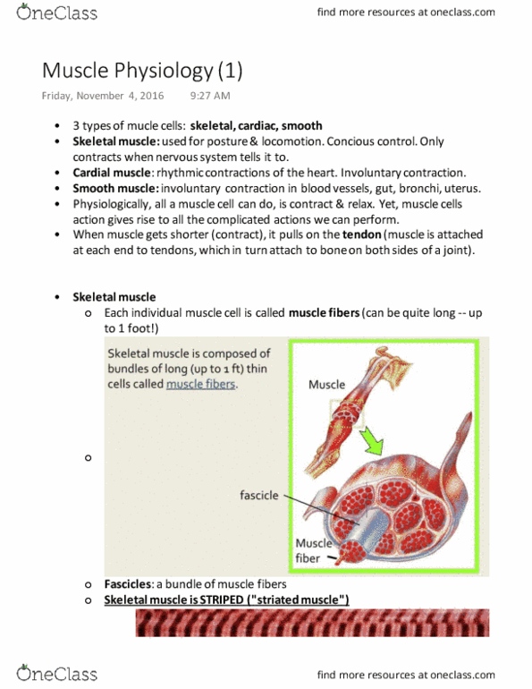
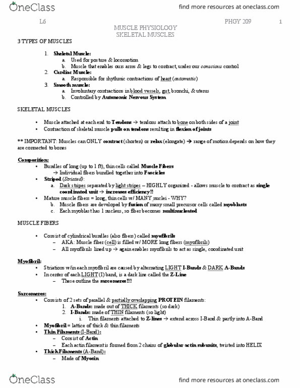
Myofibril structure: sarcomere: fundamental unit of muscle fiber, when contracts, z lines (limits) get closer together, happens along the entire length of the myofibril (get shorter, h zone is section within a band that is functionally important. Sarcomere: each sarcomere consists of two sets of parallel and overlapping protein filaments - Proteins: thin filaments made of actin --two chains of globular actin subunits twisted in a helix, thick filament made of myosin (oriented in both directions) Sliding filament model: reach out, grab, pull, let go, cycle again and again, driven by atp hydrolysis. Don"t all pull at the same time like a rowing team, but really not synchronized, act independently (at any time, some pulling) Increased calcium concentration is the signal for muscle contraction. Increments differ, one neuron can activate 10 muscle fibers (eye) or 1000s in leg. Intermediate properties: fast myosin and oxidative metabolism. Muscle response to exercise: low intensity exercise cause increase in fiber mitochondria, inceased vascularization.