PSYC 211 Lecture Notes - Lecture 29: Delta Wave, Cerebral Circulation, Muscle Tone

16 Mar 2017
School
Department
Course
Professor
Document Summary
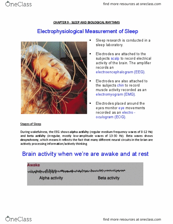
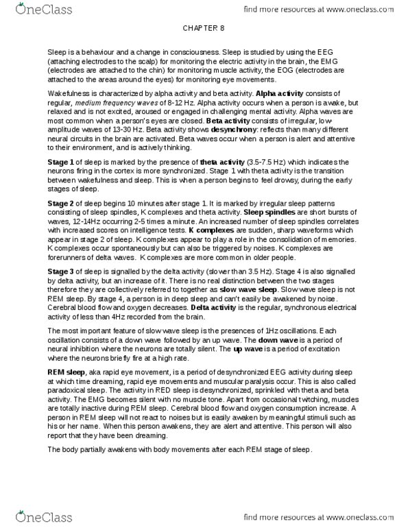
Electrophysiological measurement of sleep sleep research is conducted in a sleep lab. Electrodes are attached to the subjects scalp to record electrical activity of the brain. Electrodes are also attached to the subjects chin to record muscle activity recorded as an electromyogram (emg) Electrodes are placed around the eyes monitor eye movements recorded as an electro- oculogram (eog) Brain activity when we are awake and at rest. When we are awake, our eeg shows two basic patterns: alpha activity consists of regular, medium frequency waves (8-12 hz, beta activity is generally associated wave forms that are irregular at an amplitude of 13- Beta activity is de synchronous; it is a reflection of different neural circuits actively processing information at the same time. Theta activity: when we get drowsy, we show theta activity (3. 5-7. 5 hz). This is the transition between wakefulness and sleep. Amplitude of the eeg waves will increase and frequency becomes slower.