PSYC 212 Lecture Notes - Lecture 4: Detection Theory, Neurophysiology, Ophthalmoscopy

6 Nov 2018
School
Department
Course
Professor

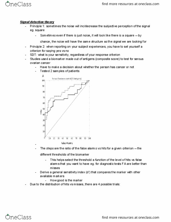
Signal detection theory
Principle #1
Sometimes the noise will increase/decrease the subjective
perception of the signal
§
Sometimes, even if there is just noise, it will look like there is a
square
§
○
Principle #2
When reporting on your subjective experiences, you have to set
yourself a criterion for saying "yes" vs "no"
§
○
Measuring the sensitivity regardless of the personal response criterion
○
•
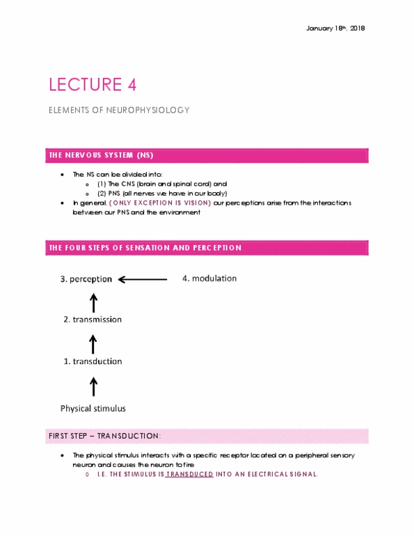
Elements of neurophysiology
Four steps of sensation and perception
Transduction
The physical stimulus interacts with a specific receptor
located on a peripheral sensory neuron and causes the
neuron to fire, i.e. the stimulus is transduced into an
electric signal
□
Doctrine of specific nerve energies
A doctrine formulated by Johannes Müller
(1801-1858) stating that the nature of a sensation
depends on which fibers are stimulated, not on how
the fibers are stimulated
®
□
1.
Transmission
The signal is transmitted to the brain
□
2.
Perception
The signal reaches the cortex and produces a conscious
perceptual experiences
□
Hardest step to explain
□
3.
Modulation
Cognitive factors, like expectations, attention, etc, will
influence how sensations are perceived
□
4.
○
•
Light and the eye
A little light physics
Light: a narrow band of electromagnetic radiation that can be
conceptualized as a wave or a stream of photons
○
Photon: a quantum of visible light (or other forms of electromagnetic
radiation) demonstrating both particle and wave properties
○
Light can be scattered, reflected, absorbed, transmitted, or refracted
Absorbed: energy (e.g. light) that is taken up and is not
transmitted at all
§
Scattered: energy that is dispersed in an irregular fashion
When light enters the atmosphere, much of it is absorbed
or scattered and never makes it to the perceiver
□
§
Reflected: energy that is redirected when it strikes a surface,
usually back to its point of origin
§
Transmitted: energy that is passed on through a surface (when it
is neither reflected nor absorbed by the surface)
§
Refracted: energy that is altered as it passes into another
medium
§
○
•
The eye - general anatomy
The human eye is made us of various parts:
Cornea The transparent "window" into the eyeball
Aqueous
humour
The watery fluid in the anterior chamber
Crystalline
lens
The lens inside the eye, which focuses light onto the
back of the eye
Pupil The dark circular opening at the center of the iris in
the eye, where the light enters the eye
Iris The coloured part of the eye, a muscular diaphragm,
that regulates light entering the eye by expanding
and contracting the pupil
Vitreous
humour
The transparent fluid that fills the large chamber in
the posterior part of the eye
Retina A light-sensitive membrane in the back of the eye
that contain rods and cones. The lens focuses an
image on the retina, which then sends signals to the
brain through the optic nerve
§
Refraction is necessary to focus light rays onto the retina and this
is accomplished by the lens
Accommodation: the process in which the lens changes its
shape, thus altering its refractive power
Nearby objects require more refraction because light
rays from near objects diverge more
®
Diopter: the focusing of a lens can be measured in
diopters, i.e. the reciprocal of the focal length
®
Focal length: distance between the lens and the point
at which light ways converge (the focus)
®
Maximal accommodation decreases with age
®
□
Problems with refractions
Emmetropia: the happy condition of no refractive
error
®
Myopia: when light is focused in front of the retina
and distant objects cannot be seen sharply;
nearsightedness
®
Hyperopia: when light is focused behind the retina
and near objects cannot be seen sharply;
farsightedness
Prebyopia: a form of hyperopia associated with
old age: eventually the lens will lose its
elasticity
◊
®
Astigmatism: unequal curving of one or more of the
refractive surfaces of the eye, usually the cornea
Multiple focal points
◊
Some of the lines won't seem to converge on
the common focal point
◊
®
□
§
○
•
The retina
Fundus: using the ophthalmoscope, doctors can view the back surface
of the patients' eyes, called the fundus
○
Optic disc: the white circle is the point where the arteries and veins
that feed the retina enter the eye, and where the axons of the ganglion
cells leave the eye towards the brain. This portion of the retina contains
no photoreceptor and is therefore "blind":
○
To experience your blind spot:
Take that figure in the textbook1.
Close your left eye2.
Look at the letter F with your right eye3.
Move the figure at approximately 15 cm from you eye4.
At some point you should see the red dot disappear… keep your
eye on the "F"
5.
○
Macula: the dark spot corresponds to the macula. It is situated in the
center of the eye (i.e. immediately behind your lens). It contains a high
density of photoreceptors and it is responsible for central vision
○
Fovea: the central part of the macula is called the fovea. It contains
almost no blood vessels, and contains a high density of photoreceptors.
It is responsible for sharp central vision
○
By contrast with other cranial nerves, the optic nerve and retina is
considered part of the brain
○
It is the only part of the central nervous system that be can imaged non
invasively
○
Can be used to diagnose certain brain disorders like Alzheimer's disease
○
Photoreceptors
Located at the back of the retina, close to the pigment
epithelium, which provides vital nutrients to the photoreceptors
§
The foremost layers of the retina are transparent
§
Transduce light energy into neural energy
§
Rods: photoreceptors specialized for night vision
Respond well in low luminance conditions□
Do not process colour□
§
Cones: photoreceptors specialized daytime vision, fine visual
activity, and colour
Respond best in high luminance conditions□
§
Much more rods (90 million) than cones (4.5 million)
§
More cones in your fovea, almost no cones outside fovea
Very poor colour vision in the periphery□
§
○
Visual angle
Vision scientists measure the size of visual stimuli by how large
an image appears on the retina, not by how large the object is
§
Rule of thumb: if you hold your thumb out at arms length, the
width of your thumbnail is about 2 degrees of visual angle
§
In summary: the visual angle of an object is a function of both its
actual size and distance from the observer
§
The foveal "rod-free" area is about 1 degree of visual angle
§
This means that we can't see much more than thumb at arm's
length in central (foveal) vision
§
○
•
Transduction of light by photoreceptors
Capturing a photon: when light hits a photoreceptor, the process of
photoactivation begins
○
Photoreceptors
Contain an outer segment (adjacent to the pigment and stored in
the outer segment
§
Pigments contain a chromophore that captures photons and a
protein, called an opsin, whose structure determines the
wavelength of light to which the photoreceptor responds
Rods have rhodopsin□
Cones have three different opsins, which respond to long,
medium, or short wavelengths
□
Some photoreceptors contain melanopsin and can monitor
ambient light levels and influence our sleep/wake cycle
□
§
○
Photoactivation: when a photon is absorbed by an opsin, it transfers its
energy to the chromophore portion of the visual pigment molecule
Once photoactivation starts, photoreceptors become
hyperpolarized (negatively charged)
Hyperpolarization is the opposite of a neuron discharging
(or depolarizing). Normally it is associated with inhibition or
neuronal activity. The fact that it is associated with the
transduction of photon is an exception here
□
1.
Changes in photoreceptor activation are communicated to the
bipolar cells in the form of graded potentials
Graded potentials vary continuously in their amplitudes□
2.
Bipolar cells synapse with retinal ganglion cells, which in an all-or-
none fashion rather than in graded potentials
3.
○
•
Lecture 4
Thursday, January 18, 2018
12:57 PM

Signal detection theory
Principle #1
Sometimes the noise will increase/decrease the subjective
perception of the signal
§
Sometimes, even if there is just noise, it will look like there is a
square
§
○
Principle #2
When reporting on your subjective experiences, you have to set
yourself a criterion for saying "yes" vs "no"
§
○
Measuring the sensitivity regardless of the personal response criterion
○
•
Elements of neurophysiology
Four steps of sensation and perception
Transduction
The physical stimulus interacts with a specific receptor
located on a peripheral sensory neuron and causes the
neuron to fire, i.e. the stimulus is transduced into an
electric signal
□
Doctrine of specific nerve energies
A doctrine formulated by Johannes Müller
(1801-1858) stating that the nature of a sensation
depends on which fibers are stimulated, not on how
the fibers are stimulated
®
□
1.
Transmission
The signal is transmitted to the brain□
2.
Perception
The signal reaches the cortex and produces a conscious
perceptual experiences
□
Hardest step to explain□
3.
Modulation
Cognitive factors, like expectations, attention, etc, will
influence how sensations are perceived
□
4.
○
•
Light and the eye
A little light physics
Light: a narrow band of electromagnetic radiation that can be
conceptualized as a wave or a stream of photons
○
Photon: a quantum of visible light (or other forms of electromagnetic
radiation) demonstrating both particle and wave properties
○
Light can be scattered, reflected, absorbed, transmitted, or refracted
Absorbed: energy (e.g. light) that is taken up and is not
transmitted at all
§
Scattered: energy that is dispersed in an irregular fashion
When light enters the atmosphere, much of it is absorbed
or scattered and never makes it to the perceiver
□
§
Reflected: energy that is redirected when it strikes a surface,
usually back to its point of origin
§
Transmitted: energy that is passed on through a surface (when it
is neither reflected nor absorbed by the surface)
§
Refracted: energy that is altered as it passes into another
medium
§
○
•
The eye - general anatomy
The human eye is made us of various parts:
Cornea
The transparent "window" into the eyeball
Aqueous
humour
The watery fluid in the anterior chamber
Crystalline
lens
The lens inside the eye, which focuses light onto the
back of the eye
Pupil
The dark circular opening at the center of the iris in
the eye, where the light enters the eye
Iris
The coloured part of the eye, a muscular diaphragm,
that regulates light entering the eye by expanding
and contracting the pupil
Vitreous
humour
The transparent fluid that fills the large chamber in
the posterior part of the eye
Retina
A light-sensitive membrane in the back of the eye
that contain rods and cones. The lens focuses an
image on the retina, which then sends signals to the
brain through the optic nerve
§
Refraction is necessary to focus light rays onto the retina and this
is accomplished by the lens
Accommodation: the process in which the lens changes its
shape, thus altering its refractive power
Nearby objects require more refraction because light
rays from near objects diverge more
®
Diopter: the focusing of a lens can be measured in
diopters, i.e. the reciprocal of the focal length
®
Focal length: distance between the lens and the point
at which light ways converge (the focus)
®
Maximal accommodation decreases with age
®
□
Problems with refractions
Emmetropia: the happy condition of no refractive
error
®
Myopia: when light is focused in front of the retina
and distant objects cannot be seen sharply;
nearsightedness
®
Hyperopia: when light is focused behind the retina
and near objects cannot be seen sharply;
farsightedness
Prebyopia: a form of hyperopia associated with
old age: eventually the lens will lose its
elasticity
◊
®
Astigmatism: unequal curving of one or more of the
refractive surfaces of the eye, usually the cornea
Multiple focal points
◊
Some of the lines won't seem to converge on
the common focal point
◊
®
□
§
○
•
The retina
Fundus: using the ophthalmoscope, doctors can view the back surface
of the patients' eyes, called the fundus
○
Optic disc: the white circle is the point where the arteries and veins
that feed the retina enter the eye, and where the axons of the ganglion
cells leave the eye towards the brain. This portion of the retina contains
no photoreceptor and is therefore "blind":
○
To experience your blind spot:
Take that figure in the textbook1.
Close your left eye2.
Look at the letter F with your right eye3.
Move the figure at approximately 15 cm from you eye4.
At some point you should see the red dot disappear… keep your
eye on the "F"
5.
○
Macula: the dark spot corresponds to the macula. It is situated in the
center of the eye (i.e. immediately behind your lens). It contains a high
density of photoreceptors and it is responsible for central vision
○
Fovea: the central part of the macula is called the fovea. It contains
almost no blood vessels, and contains a high density of photoreceptors.
It is responsible for sharp central vision
○
By contrast with other cranial nerves, the optic nerve and retina is
considered part of the brain
○
It is the only part of the central nervous system that be can imaged non
invasively
○
Can be used to diagnose certain brain disorders like Alzheimer's disease
○
Photoreceptors
Located at the back of the retina, close to the pigment
epithelium, which provides vital nutrients to the photoreceptors
§
The foremost layers of the retina are transparent
§
Transduce light energy into neural energy
§
Rods: photoreceptors specialized for night vision
Respond well in low luminance conditions□
Do not process colour□
§
Cones: photoreceptors specialized daytime vision, fine visual
activity, and colour
Respond best in high luminance conditions□
§
Much more rods (90 million) than cones (4.5 million)
§
More cones in your fovea, almost no cones outside fovea
Very poor colour vision in the periphery□
§
○
Visual angle
Vision scientists measure the size of visual stimuli by how large
an image appears on the retina, not by how large the object is
§
Rule of thumb: if you hold your thumb out at arms length, the
width of your thumbnail is about 2 degrees of visual angle
§
In summary: the visual angle of an object is a function of both its
actual size and distance from the observer
§
The foveal "rod-free" area is about 1 degree of visual angle
§
This means that we can't see much more than thumb at arm's
length in central (foveal) vision
§
○
•
Transduction of light by photoreceptors
Capturing a photon: when light hits a photoreceptor, the process of
photoactivation begins
○
Photoreceptors
Contain an outer segment (adjacent to the pigment and stored in
the outer segment
§
Pigments contain a chromophore that captures photons and a
protein, called an opsin, whose structure determines the
wavelength of light to which the photoreceptor responds
Rods have rhodopsin□
Cones have three different opsins, which respond to long,
medium, or short wavelengths
□
Some photoreceptors contain melanopsin and can monitor
ambient light levels and influence our sleep/wake cycle
□
§
○
Photoactivation: when a photon is absorbed by an opsin, it transfers its
energy to the chromophore portion of the visual pigment molecule
Once photoactivation starts, photoreceptors become
hyperpolarized (negatively charged)
Hyperpolarization is the opposite of a neuron discharging
(or depolarizing). Normally it is associated with inhibition or
neuronal activity. The fact that it is associated with the
transduction of photon is an exception here
□
1.
Changes in photoreceptor activation are communicated to the
bipolar cells in the form of graded potentials
Graded potentials vary continuously in their amplitudes□
2.
Bipolar cells synapse with retinal ganglion cells, which in an all-or-
none fashion rather than in graded potentials
3.
○
•
Lecture 4
Thursday, January 18, 2018 12:57 PM
Document Summary
Sometimes the noise will increase/decrease the subjective perception of the signal. Sometimes, even if there is just noise, it will look like there is a square. When reporting on your subjective experiences, you have to set yourself a criterion for saying yes vs no Measuring the sensitivity regardless of the personal response criterion. The physical stimulus interacts with a specific receptor located on a peripheral sensory neuron and causes the neuron to fire, i. e. the stimulus is transduced into an electric signal. A doctrine formulated by johannes m ller (1801-1858) stating that the nature of a sensation depends on which fibers are stimulated, not on how the fibers are stimulated. The signal reaches the cortex and produces a conscious perceptual experiences. Cognitive factors, like expectations, attention, etc, will influence how sensations are perceived. Light: a narrow band of electromagnetic radiation that can be conceptualized as a wave or a stream of photons.