PSYC 311 Lecture Notes - Lecture 2: Medial Frontal Gyrus, Lateral Sulcus, Inferior Temporal Gyrus

8 Jun 2018
School
Department
Course
Professor

Tuesday, May 2nd, 2017
1
PSYC 311
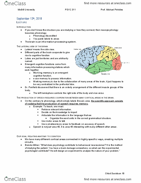
Week 1 Lecture 2: Anatomy of the Brain
Review from last lecture
- Dr. Penfield was a neuro surgeon focusing on epilepsy
o Patients that cannot be treated with medicine, will have part of their brain
removed
- Brenda Milner (from McGill)
o She was an experimental psychology, trained to evaluate cognition: memory,
perception, learning, and spatial ability
o eoe pat of patiets’ ai, ad the evaluate patients before and after
surgery, see if the operation was worth it.
o It is the beginning of psychologists, people are trained to measure cognition and
perception
o She became famous because of patient H.M
▪ Not operated in Montreal, in the States, but because Brenda Milner was
well-known, she has a chance to study the patient
▪ H.M’s hippocampus was removed bilateral
• He ould’t lea
• He remembered anything before
Lecture content:
- Neuropsychology:
o Cannot be taught unless they know enough about structure of the brain.
- Left hemisphere: Verbal processes
- Right hemisphere: visual spacial ability, understanding/ remembering faces: Non- verbal
process
- Investigate on what happens when you separate two hemispheres:
o Sometimes disease will cause separation of two hemispheres
o Sometimes in patients with severe epilepsy, couldn't be controlled from
medicine, will have to get a surgery to separate the two hemispheres
- Primates’ Brian:
o Highly gyrated: cerebral cortex: gray matter of the surface of the brain
▪ Different from cats and rats.. We have more cortex
▪ The rest of the brain: subcortical: this is the part that is very similar to
cats and rats
• Regulate breathing
• Motivation
• Rewards
o In primates, cerebral cortex has expended, so it has to be folded to fit into the
brain.
o Cortex within the folds: sulcus
o Between the folds: gyrus
o When we study the surface anatomy: we are going to understand various folds
find more resources at oneclass.com
find more resources at oneclass.com

Tuesday, May 2nd, 2017
2
Morphological:
Lateral surface (review from last lecture) :
1. Central sulcus:
- Dr. Penfield mapped the brain through stimulation.
- M1: pre-central gyrus+ half sulci
o Since sulci is folded inwards, half of the folded area, along with the pre-central
gyrus controls motor movements
- M1: motor movements
o Getting muscle twitch of the opposite side of the body
o From top to bottom: Leg, arm, face
- 50% of cortex is not on surface, in sulci.
- S1: posterior part: somatosensory movements
o Sensation: sense of touch
- Penfield established two concepts for M1 and S1:
1. Somatotopic representation: In many other parts of the body/brain, we have orderly
representation of muscles. Soma: body / totopic: place
o Each part of the body has its place in the brain
▪ The legs: at the top
▪ The hand
▪ The face
o They are all really nicely represented.
2. Contralateral representation: brain hemisphere control of opposite of the body
o Left hemisphere controls right side of the body
o Right hemisphere controls left side of the body
- Central sulcus divides frontal cortex and parietal cortex:
o Parietal cortex: spacial information
o Frontal cortex: organize/ plan activities.
Lateral fissure/ Sulcus sulci:
- Fissure: big sulci, have sulcus in fissure
Intra- parietal sulcus:
- Separates Superior lobule and inferior lobule
Superior temporal sulcus
- Superior temporal gyrus:
o left hemisphere: voices convey words (verbal)
o Right hemisphere: songs that convey music
o Primary auditory cortex: hidden in the sulcus of the lateral fissure (?)
o Heschl Gyrus: primary auditory cortex: Where is the auditory info first comes to
cortex
find more resources at oneclass.com
find more resources at oneclass.com
Document Summary
Week 1 lecture 2: anatomy of the brain. Dr. penfield was a neuro surgeon focusing on epilepsy: patients that cannot be treated with medicine, will have part of their brain removed. Neuropsychology: cannot be taught unless they know enough about structure of the brain. Right hemisphere: visual spacial ability, understanding/ remembering faces: non- verbal process. Lateral surface (review from last lecture) : central sulcus: Dr. penfield mapped the brain through stimulation. M1: pre-central gyrus+ half sulci: since sulci is folded inwards, half of the folded area, along with the pre-central gyrus controls motor movements. M1: motor movements: getting muscle twitch of the opposite side of the body, from top to bottom: leg, arm, face. 50% of cortex is not on surface, in sulci. S1: posterior part: somatosensory movements: sensation: sense of touch. Penfield established two concepts for m1 and s1: somatotopic representation: in many other parts of the body/brain, we have orderly representation of muscles.