PSYC 427 Lecture Notes - Lecture 10: Capuchin Monkey, Posterior Parietal Cortex, Premotor Cortex

14 May 2018
School
Department
Course
Professor

PSYC 427 – LECTURE 10
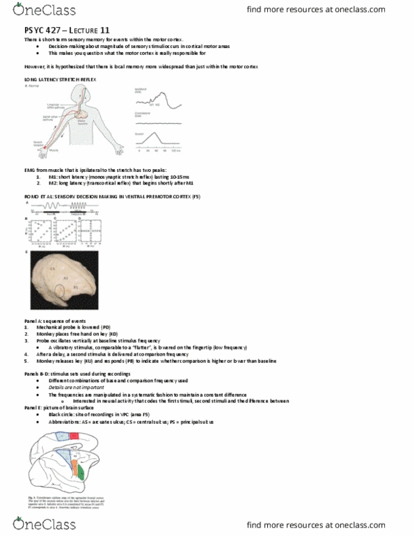
Single cells in monkey motor cortex are activated at short-latency by deep proprioceptive and cutaneous stimuli
• Short latency sensory input reaches M1 from somatosensory cortex, and possibly directly from the thalamus
• Proprioceptive: change in joint position
• Short latency, or transcortical, reflexes are quite adaptive- more specifically, they are sensitive to the nature of the task that the animal has to perform.
The motor cortex has sensory receptive fields that respond to sensory input, mostly via the somatosensory cortex.
• The motor cortex seems to involve more than motor responses (may be sensitive to more general cognitive functions).
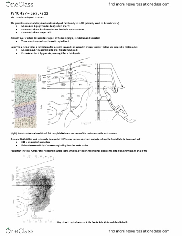
ANATOMY: INPUTS INTO PRIMARY MOTOR CORTEX
• The location and extent of the injection cores in M1 are outlines with large color-coded areas with black contours.
o Retrograde tracer was injected in different locations in different monkeys to rule out observations being due to tracer location
• Labelled cells are shown with small dots that are colored according to the same colors used for injection cores
• Boundaries in parietal cortex are defined cytoarchitectonically
Nua’s ok o eus okes as iflueed eletophsiologial tehiues leaed fo Nudo
• Questioned where motor inputs originate from
• Cebus are new world monkeys with individuated control of digits
o Able to recognize themselves in mirrors (different reaction when reflection is of another monkey and not the self)
o Has new motor cortex
There is remarkable segregation in the motor cortex: rostral areas mostly get inputs from other cortical motor areas, whereas the more caudal areas mostly get
inputs from somatosensory areas.
• Caudal to the central sulcus: areas 1,2, and 5 (posterior parietal cortex)
• Area 3, part of the somatosensory cortex that is buried within the central sulcus, was not recorded from
The parietal operculum, underneath the lateral sulcus, contains the second somatosensory area
• Similar to the opening of an eyelid
Majority of red and green cells are projecting to the injection site just ahead of the central sulcus
• In other words, somatosensory neurons are projecting to the primary motor cortex
find more resources at oneclass.com
find more resources at oneclass.com

PMv (ventral premotor cortex) contains largely hand-area cells (digit movement)
• Projects to more rostral and lateral area of the primary motor cortex
PMd (dorsal premotor cortex) contains largely arm-area cells
• Projects to more rostral and medial area of the primary motor cortex
There is almost no anatomical somatosensory input to these areas.
Caudo-medial region, just rostral to the central cortex, is the new motor cortex
• Inputs mostly from the parietal cortex (area 5), though there is some distribution
Caudo-lateral region
• Inputs almost entirely from sensory areas of the brain
The proportion of labeled cells in a distant cortical area was divided by that of the area with the most labeled cells.
• For example, for RM injections, PMd had the highest proportion of labeled cells (normalized value = 1.0)
o Other areas are relative to largest possible input. For example, in the case of RL cells, 0.35 are from the PMd relative to 1.0 from the PMv
• The percentage of labeled cells found in SMA, following RM injections, was approximately 4x lower than in PMd (normalized value = 0.26)
o SMA and somatosensory areas are not contributing much to the old motor cortex
o More rostral areas are receiving inputs from the motor cortex. On the other hand, more caudal areas are receiving inputs from the primary
somatosensory cortex
B panel: summarizes pattern of projections to the M1 hand representation from the different areas of the sensorimotor cortex
• Density of the projections is represented as line thickness
There is anatomical segregation of input into the motor cortex (keep in mind that this is in cebus monkeys): vast majority of inputs into rostral motor cortex come
from motor areas; vast majority of inputs into caudal motor cortex come from sensory areas.
Like neurons in somatosensory cortex, neurons in motor cortex have sensory receptive fields. Some respond to cutaneous stimuli, others to muscle and joint
afferent input.
Murphy recorded (extracellular) a large population of single units from precentral forelimb areas of awake monkeys
• Single units were activated by cutaneous input (C), joint motion (J) or both.
o Cutaneous: skin contact
o Joint rotation: at the level of the wrist
find more resources at oneclass.com
find more resources at oneclass.com
Document Summary
Single cells in monkey motor cortex are activated at short-latency by deep proprioceptive and cutaneous stimuli. Short latency sensory input reaches m1 from somatosensory cortex, and possibly directly from the thalamus. Short latency, or transcortical, reflexes are quite adaptive- more specifically, they are sensitive to the nature of the task that the animal has to perform. The motor cortex has sensory receptive fields that respond to sensory input, mostly via the somatosensory cortex. The motor cortex seems to involve more than motor responses (may be sensitive to more general cognitive functions). The location and extent of the injection cores in m1 are outlines with large color-coded areas with black contours. Retrograde tracer was injected in different locations in different monkeys to rule out observations being due to tracer location. Labelled cells are shown with small dots that are colored according to the same colors used for injection cores.