PSYC 427 Lecture Notes - Lecture 13: Premotor Cortex, Frontal Lobe, Anterograde Tracing

Document Summary
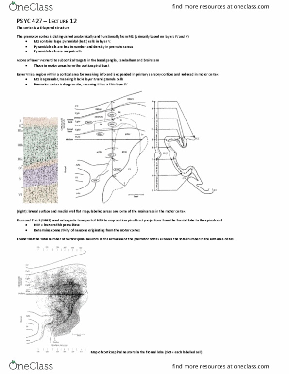
Figure shows motor areas on the lateral surface of the frontal lobe. Premotor cortex is distinguishable anatomically and functionally from m1. M1 contains large pyramidal cells (betz cells) in layer v output cells. Density of pyramidal cells less in premotor areas and smaller. M1 is agranular, lacks layer iv (granule cells). In figure on top, can see somatotopic organization of body in central sulcus and up on the precentral gyrus. Identified on the basis of difference in density of granule cells. J panel increase discharge during comparison period when second stimulus is lower frequency and terminate activity when second stimulus is higher in frequency. G, j aspects of second stimulus in relation to first. Firing rate modulation as a function of the base. Sma has extensive inputs and outputs. presma gets input/output from prefrontal areas. As stimulus frequency increases of vibration, cell firing rate increases. So this cell has the capacity to code information about the stimulus.