BIOLOGY 2B03 Lecture Notes - Lecture 2: Chronic Obstructive Pulmonary Disease, Methylation, Sickle-Cell Disease

6 Jun 2018
School
Department
Course
Professor

From Polypeptide to Protein: Protein Folding
Secondary Structure
conformation of a portion of polypeptide •
Motifs: combinations of secondary structures ◦
periodic folding of the polypeptide chain into distinct, conserved, geometric arrangements •
alpha-helix: spiral, rod-like structure ◦
beta-sheet: planar structure, composed of alignments of 2 or more B strands ◦
turns and loops: connectors ◦
R groups not involved with structure •
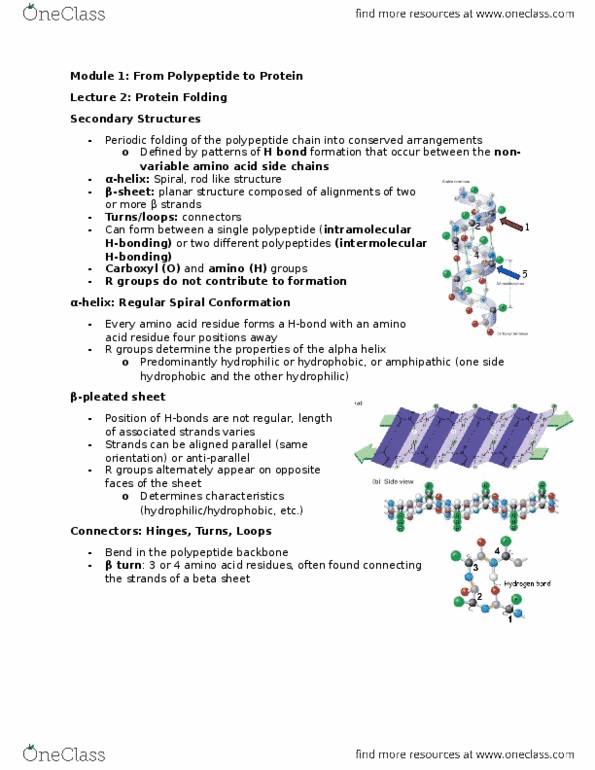
Alpha-Helix
regular spiral conformation •
carbonyl oxygen of each peptide bond is H-bonded to the amide hydrogen of the ◦
amino acid four residues toward C-terminus
forms independently of specific amino acid side chains ◦
a cylinder with side chains pointing out •
R groups determine hydrophobic/hydrophilic quality of the outer surface of the ◦
helix
Beta-Pleated Sheet
laterally packed B strands (5-8 AA residues long) H-bonds •
between carboxyl and amino groups of backbone in adjacent B
strands
R groups determine hydrophobic/hydrophilic quality of the ◦
surfaces of sheets
antiparallel and parallel sheets •
Connectors: Hinges, Turns Loops
these are ways the polypeptide backbone bends •
on the right is an image of a beta turn •
Motifs
secondary structures organized into motifs •
motifs are built from specific combinations of secondary structures ◦
motifs are structural units that recur in many proteins •
have 3D architecture •
usually associated with specific function •
Motif Examples
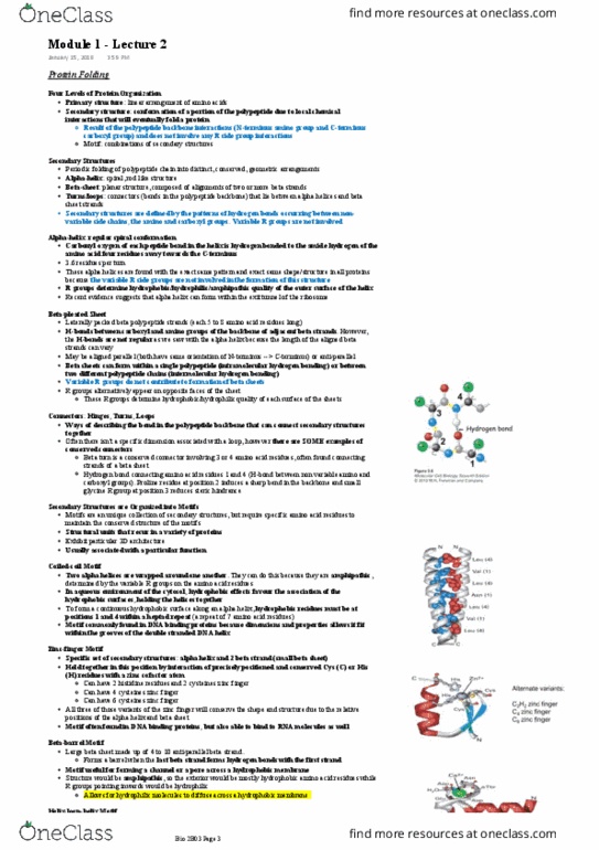
Coiled-coil: two alpha helices wrapped around one another •
hydrophobic surfaces spiral around each other ◦
Zinc-finger: consists of an alpha-helix and 2 beta-strands that are •
held in position by the interaction of Cysteine or Histidine residues with a zine atom
can bind to RNA ◦
B-barrel: a beta-sheet forms a barrel when the last strand forms hydrogen bonds with the •
first strand
useful for creating channel/pore across membrane ◦
4-10 antiparallel B-strands form a sheet ◦
Helix-loop-helix: two alpha helixes joined by a loop region •
loop region can bind to Ca2+ (cofactor) via carboxyl side chains from Asp or Glu ◦
in loop
Document Summary
Secondary structure conformation of a portion of polypeptide. Motifs: combinations of secondary structures periodic folding of the polypeptide chain into distinct, conserved, geometric arrangements alpha-helix: spiral, rod-like structure beta-sheet: planar structure, composed of alignments of 2 or more b strands turns and loops: connectors. Alpha-helix regular spiral conformation carbonyl oxygen of each peptide bond is h-bonded to the amide hydrogen of the amino acid four residues toward c-terminus forms independently of specific amino acid side chains a cylinder with side chains pointing out. R groups determine hydrophobic/hydrophilic quality of the outer surface of the helix. Beta-pleated sheet laterally packed b strands (5-8 aa residues long) h-bonds between carboxyl and amino groups of backbone in adjacent b strands. R groups determine hydrophobic/hydrophilic quality of the surfaces of sheets antiparallel and parallel sheets. Coiled-coil: two alpha helices wrapped around one another hydrophobic surfaces spiral around each other.