BIOLOGY 2B03 Lecture Notes - Lecture 22: Histone H3, Histone H1, Spindle Apparatus

18 Jun 2018
School
Department
Course
Professor

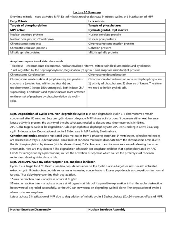
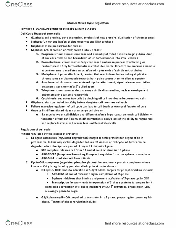
Cell Cycle Dynamics and Checkpoints: Targets of MPF
Entry Into Mitosis Requires Active MPF
phosphorylation mediated by active MPF complex - a heterodimer of mitotic Cyclin and CDK •
all changes in cells during mitosis (chromosomes condensing, nuclear envelope break, etc.) must be reversed •
after mitosis
this is done by inactivation of MPF through ubiquitin mediated depredated of Cyclin B ◦
cytosolic phosphatases reverse phosphorylation of same key proteins that initiate these events ◦
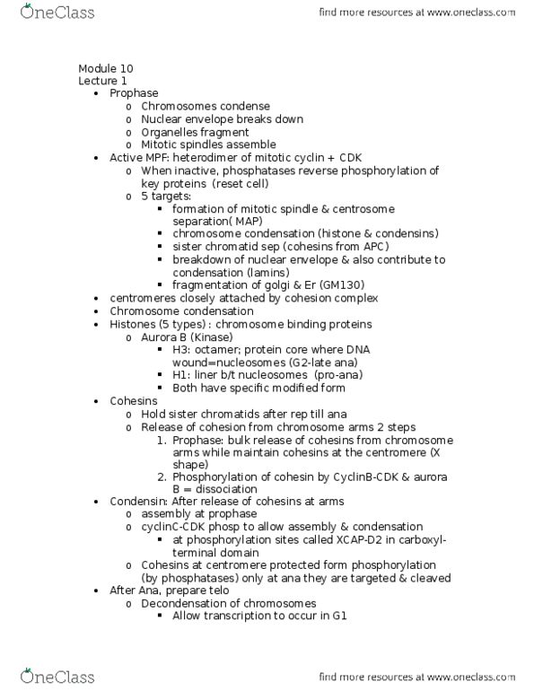
Active MPF Kinase: Prophase Events
following processes initiated by MPF-mediated phosphorylation of target proteins at entry into mitosis •
1) formation of mitotic spindle through phosphorylation of microtubule-associated proteins promoting ◦
instability and centrosome separation
2) condensation of chromosomes through phosphorylation of condensing and histone proteins ◦
3) preparation for sister chromatid separation through phosphorylation of cohesins ◦
4) breakdown of nuclear envelope through phosphorylation of nuclear lamins ◦
5) fragmentation of Golgi and ER via phosphorylation of GM130 ◦
Inactivation of MPF: Telophase Events
progression through mitosis + exit requires decrease in mitotic cyclins and inactivation of MPF •
also requires activity of phosphatases to dephosphorylate MPF target proteins ◦
inactivation of MPF allows for: •
nuclear envelope reassembly ◦
chromosome decondensation ◦
mitotic spindle disassembly ◦
endomembrane system reusables ◦
Chromosome Condensation
highly compact chromosomes in have centromeres that are closely attached by cohesion complexes while arms •
have separated apart from on another slightly
targets of MPF: •
condensins ◦
cohesins ◦
histones, H1 and H3 ◦
topoisomerase ◦
Histones
many proteins for chromosome condensation and phosphorylation is key regulatory switch in each case •
DNA organized around chromosomal binding proteins called histones •
5 types of histones: ◦
histone H3 - part of octamer that forms protein for around which DNA is wound to create nucleosome ‣
histone H1 - linker between neighbouring nucleosomes ‣
H1 + H3 phosphorylated by kinase Aurora B during condensation •
Cohesins
2 other protein families important to mitotic chromosome organization - cohesins and condensins •
cohesin proteins form cohesin complex - required to hold sister chromatids together after replication •
release of cohesins = 2 steps: ◦
1) bulk release of cohesins from chromosome arms while maintaining cohesins at centromere ‣
this produces the X-shaped chromosome we always see •
2) phosphorylation of cohesin subunits by multiple kinases, including CyclinB-CDK and Aurora B allows ‣
dissociation to occur