KINESIOL 1AA3 Lecture Notes - Lecture 20: Renal Corpuscle, Connective Tissue, Renal Calyx

3 Apr 2016
School
Department
Course
Professor
Document Summary
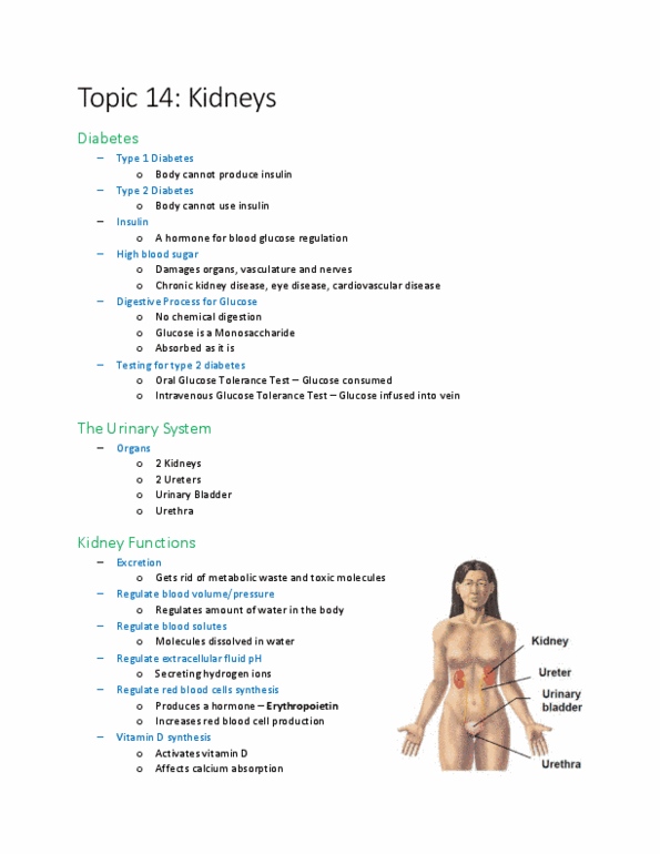
>10 million people inc canada have it. Blood is sampled of 3 hr period. Check the time around 1 (when glucose started to get absorbed) and 2 hours ( when glucose is all absorbed) Urethra (1: urine flow: kidneys >> ureters >> bladder >> urethra. Regulation of red blood cell synthesis: produce hormone (epo) which increases production of rbc: vitamin d synthesis: activate vitamin d (effects ca2+ levels) External anatomy of kidney: bean-shaped organ, size of clenched fist. Sunday, april 3, y: ~130 g, retroperitoneal, posterior abdominal wall, lateral to vertebral column, above the waist, left kidney is relatively superior because. Liver is superior to right kidney: renal capsule, fibrous connective tissue surrounding kidney, adipose issue ( perirenal , mechanical shock absorber, renal fascia, thin connective tissue, anchor to surrounding tissue, adipose tissue ( pararenal ) Renal vein and ureters exit: renal sinus. Two regions: medulla (inner): renal pyramids (~10-20, cortex (outer): renal columns (space between pyramids)