KINESIOL 1AA3 Lecture Notes - Lecture 3: Cardiac Skeleton, Skeletal Muscle, Myofibril

6 Jul 2016
School
Department
Course
Professor
Document Summary
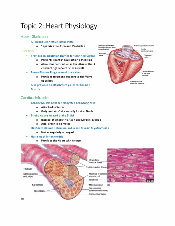
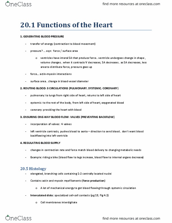
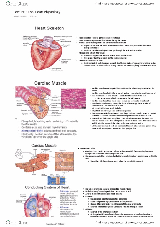
Fibrous connective tissue plate that separates the aria and ventricles (rings), support the valves. Provides an attachment point for the ventricular muscles, contracting along length. Contracts differently than skeletal muscle, apex moves towards the skeleton, twisting motion (wringing wet clothes motion) effectively pumps blood through valves. Other main job of skeleton is to provide a barrier betw electrical signals in atria and vetricles. In heart, when one cell is stimulated, the whole organ will contract, the skeleton helps have contraction in specific parts (control the signal) Elongated, branching cells (shorter than skeletal muscle) containing 1-2 centrally located nuclei. Contains actin and myosin myofilaments, not as many myofibrils as skeletal muscle. Striated banding, many mitochondria ( needs a constant supply of atp due to the constant use of the heart) T-tubules are wider in diameter than skeletal, wrap around myofibrils at z-disk, less efficient. Intercalated disks: connection point between 2 neighboring cardiac cells, finger-like projections fit together and give more stability.