MEDRADSC 2I03 Lecture Notes - Lecture 10: Airway Obstruction, Mycobacterium Tuberculosis, Bacterial Pneumonia

21 Oct 2016
School
Department
Course
Professor
Document Summary
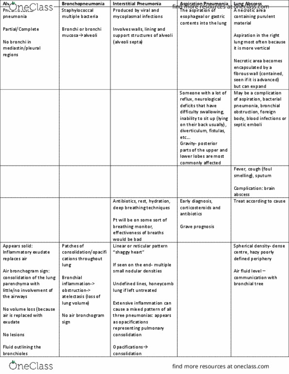
Can involve pulmonary segments or entire lobe. No bronchi in mediastinal or pleural regions- r/o lesion. Walls, lining and support structure of alveoli. Extensive inflammation- mixed pattern of three pneumonias. Pt will be on some sort of breathing monitor, effectiveness of breaths would be bad. Esophageal or gastric contents: someone with a lot of reflux, neurological deficits that have difficulty swallowing, inability to sit up (lying o(cid:374) their (cid:271)a(cid:272)k usually), diverti(cid:272)ulu(cid:373), fistulas, et(cid:272) . Posterior upper and lower lobes of the lungs. Bacterial pneumonia, bronchial obstruction, foreign body, blood infections or septic emobli. Encapsulated (contained, seen if it is advanced) but can expand. Spherical density- dense centre, hazy poorly defined periphery. Air fluid level communication with bronchial tree. Black arrow: pus like purulent in the cavity. Lungs affected (shows up in apical portion of lungs first, hard to see and as it progresses it can affect- gi, gu and skeletal. Number of bacilli and resistance of infected tissue.