MEDRADSC 2I03 Lecture Notes - Lecture 12: Infant Respiratory Distress Syndrome, Pleural Effusion, Pulmonary Edema

30 Nov 2016
School
Department
Course
Professor
Document Summary
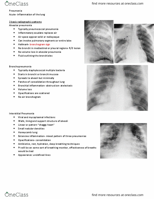
Edema: accumulation of fluids in intercellular spaces or body cavities. Sinusitis (maxillary, ethmoid, frontal, sphenoid are lined with nasal mucosa) Viral infection of upper respiratory tract can block drainage of the sinuses. Usually result of spread of adjacent infection (surgery, trauma, instrumentati on in pleural space) In a pt with chf an effusion may develop in an interlobar fissure, appearing as a round density (mimics solitary pulmonary nodule) Rupture of subpleural bulla (spontaneously or b/c of emphysema) B/c of neonatal hyaline membrane disease (lack of surfactant) Large: may need prompt chest tube drainage/suction to remove the air. Antibiotics, decongestants, steroid nasal spray (if chronic): surgery to clean or repair obstruction. Sinus tract or fistula may require drainage tube placement. No vascular markings on the right side= lung collapse. A: arrowheads pointing to pleural line (small pneumothorax there) No vasculature on the outside of the arrows (apical pneumothorax)