MEDRADSC 2I03 Lecture 12: Respiratory Pathologies

11 Dec 2018
School
Department
Course
Professor
Document Summary
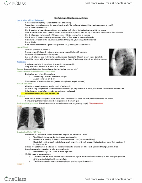
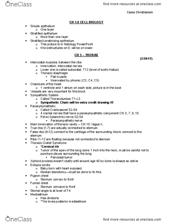
How to view a chest radiograph: trace diaphragm. Normal chest x-ray - costal phrenic angle + mediastinum. Same density -> should see air here, if not -> pathology. Should see costal phrenic angle: check size/shape of heart, check position of heart & mediastinum. Widening -> fluid collection: check hilum. Dense -> fluid accumulation: check lungs. If v pronounced -> fluid within: ribs/clavicle/rotation. Medial ends of clavicle over vertebrae -> indicates rotation: count 10 ribs. Indicates good rotation: superimposition of ribs posterior to vertebrae. If rotation -> affects what can be seen: normal space in front and behind -> if not, indicates pathology, open thoracic intervertebral disc spaces, note: upper retrosternal area and retrocardiac area = same density. Atelectasis - collapsed lung: most common result of brachial obstruction. When tube is not positioned properly: diminished air, reduced lung volume. Airless lung - plate like streaks to collapse.