ANAT 100 Lecture Notes - Lecture 1: Mononuclear Phagocyte System, Hepatic Artery Proper, Cholecystokinin

9 Dec 2015
School
Department
Course
Professor
Document Summary
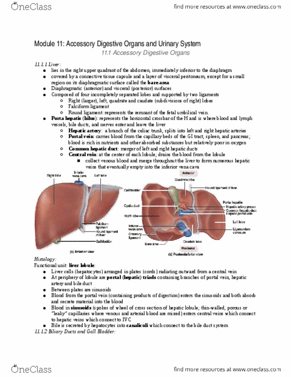
Composed of four incompletely separated lobes and supported by two ligaments. Major lobes are the left and right lobes. The right lobe is separated from the smaller left lobe by the falciform ligament, a peritoneal fold that secures the liver to the anterior abdominal wall. Round ligament of the liver lies in the inferior free edge of the falciform ligament (remnant of the fetal umbilical cord). Subdivisions of the right lobe include the caudate lobe and the quadrate lobe. Along the surface of the liver are several structures that collectively resemble the letter h; Porta heptis represents the horizontal crossbar of the h and is where blood and lymph. Inferior vena cava and ligamentum venosum form the vertical inferior parts. vessels, bile ducts and nerves enter the leave the liver. Connective tissue capsule branches through the liver and forms septa that partition the liver into thousands of small, polyhedral hepatic lobules.