ANAT 216 Lecture Notes - Lecture 8: Simple Squamous Epithelium, Juxtaglomerular Cell, Podocyte

13 Oct 2016
School
Department
Course
Professor
Document Summary
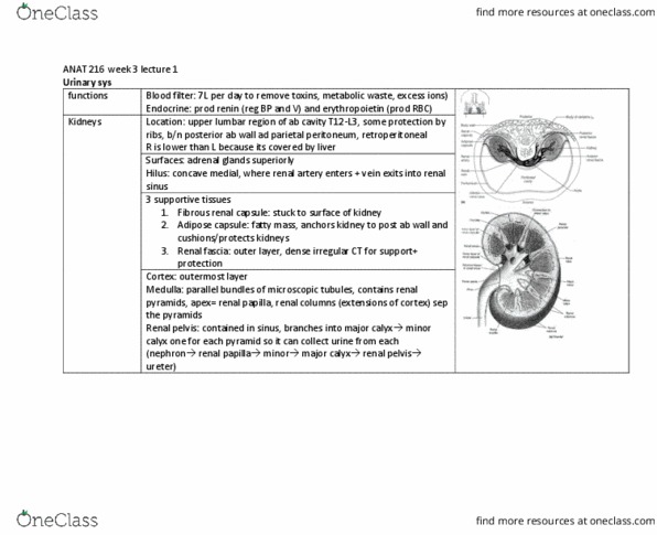
Urinary system: lecture 1 (reading material: chapter 26: pages 695-706) Major excretory organs of body: unappreciated until malfunctioned. Slightly covered by ribs: covered by liver right side sits lower, size of large bar of soap* lateral surface is smooth, medial surface is concave. Where renal artery enters artery and renal vein leaves. Gross anatomy: location: size, surface anatomy: lateral suface is smooth. Cleft on medial side: renal sinus. Space inside kidney open to hilus: supportive tissues, renal capsule fibrous. Penetrate adipose capsule to join renal fascia: causes suspension of kidneys, adipose capsule. Internal anatomy: cortex, medulla, pelvis, cortex, outermost layer in contact with renal capsule, medulla, middle layer, medulla or renal pyramids: Parallel bundles of microscopic tubules causing stripes: sinus filled with hilus filled with fat, renal papilla. Apex of renal pyramid: renal columns. Extensions of cortical tissue between medulla pyramids lobes. Each kidney has 6 to 18 lobes: renal pelvis: lateral to hilus within sinus flat funnel shaped tube.