ANAT 216 Lecture Notes - Lecture 15: Cystic Duct, Exocrine Gland, Muscular Layer

13 Oct 2016
School
Department
Course
Professor
Document Summary
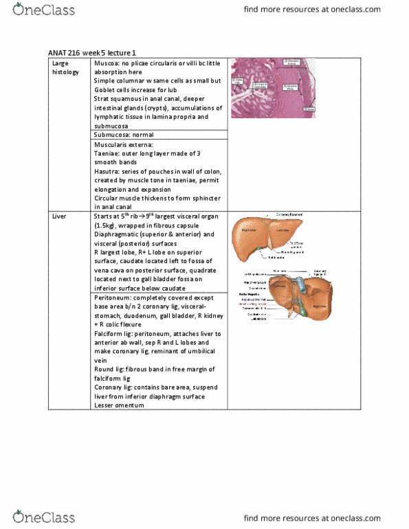
Digestive system: lecture 4 (reading material: chapter 25: pages 682-689) Liver: diaphragmatic (anterior, visceral (posterior) surfaces, right, left, quadrate and caudate lobes. On right side but projects to the left. Quadrate inferior, posterior: 1. 5 kg largest internal organ, peritoneum: interperitoneal falciform ligament: round ligament ligamentum teres remnence of fetal vein that brough blood to body from placenta to abdominal wall from liver. Atattches to diaphragm lesser omentum: porta hepatis (hilus): Hepatic artery (proper): branch of celiac artery, carries oxygenated blood to the liver tissues (hepatic) portal vein, carries nutrient-rich venous blood from the digestive tract, from splenic vein, superior and inferior mesentery veins. Bile duct: carries bile from the liver, hepatic veins: take venous blood from the liver to the ivc, histology: functional unit: liver lobule: Hexagonal shape lobule liver cells (hepatocytes) arranged in plates (cords) radiating outward from a central vein (spokes of a wheel) lines of cells: between are sinusoidal capillaries.