ANAT 216 Lecture Notes - Lecture 4: Pleural Cavity, Lamina Propria, Bronchus

28 Apr 2018
School
Department
Course
Professor

ANAT 216 week 2 lecture2
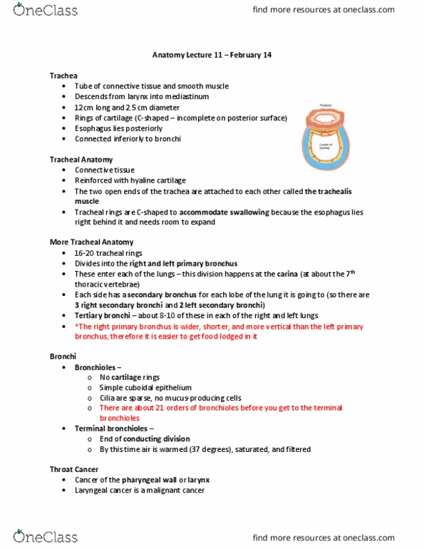
Trachea
Wind pipe, 10-12cm length and 2.5cmD, divides into primary bronchi, 3 layers
Mucosa: lines lumen, RTE supported by lamina propria
Submucosa: layer of dense irregular CT w mucous glands
Adventitia: outermost layer, reinforced by 13-20 C shaped cartilage rings to keep
passageway open, aerolar CT
Trachealis: layer of smooth muscle at posterior so bolus of food can pass easily,
contraction decreases D of lumen to change velocity of air flow
Carina: last cart ring, marks where trachea ends and primary bronchi begins
Primary
bronchi
Start at T5
Hilus: groove, only entry/exit for structures associated w bronchi, roots of the lung
R bronchi is smaller, shorter and more vertical so most objects will get stuck here
Lungs
Pleural
memb
Parietal: outer, covers top of diaphragm, projects inb/n lung and heart,
fuses w pericardium forming lateral wall of mediastinum
Visceral: tightly bound to lungs
Pleural cavity: b/n two memb, pleural fluid acts as lubricant for thoracic
wall and lungs
Surfaces
Apex/ cupola: superior tip, extends to base of neck above level of 1st rib
Diaphragmatic: concave surface that rests on diaphragm
Coastal: curving surface distal
Mediastinal:
- Hilum: groove where airways, arteries and nerves pass to get
deeper in/out of lungs (root of the lung)
- Membranous + structural comp:
R vs L lung
Left: groove for aorta and associated vessels, cardiac notch forming
tongue (pneumonia site), longer, grooves only seen post-mortem
find more resources at oneclass.com
find more resources at oneclass.com
Document Summary
Wind pipe, 10-12cm length and 2. 5cmd, divides into primary bronchi, 3 layers. Mucosa: lines lumen, rte supported by lamina propria. Submucosa: layer of dense irregular ct w mucous glands. Adventitia: outermost layer, reinforced by 13-20 c shaped cartilage rings to keep passageway open, aerolar ct. Trachealis: layer of smooth muscle at posterior so bolus of food can pass easily, contraction decreases d of lumen to change velocity of air flow. Carina: last cart ring, marks where trachea ends and primary bronchi begins. Hilus: groove, only entry/exit for structures associated w bronchi, roots of the lung. R bronchi is smaller, shorter and more vertical so most objects will get stuck here. Parietal: outer, covers top of diaphragm, projects inb/n lung and heart, fuses w pericardium forming lateral wall of mediastinum. Pleural cavity: b/n two memb, pleural fluid acts as lubricant for thoracic wall and lungs. Apex/ cupola: superior tip, extends to base of neck above level of 1st rib.