ANAT 216 Lecture Notes - Lecture 11: Superior Mesenteric Vein, Superior Mesenteric Artery, Simple Columnar Epithelium

28 Apr 2016
School
Department
Course
Professor
Document Summary
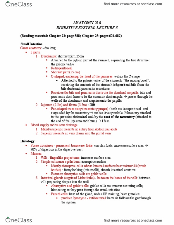
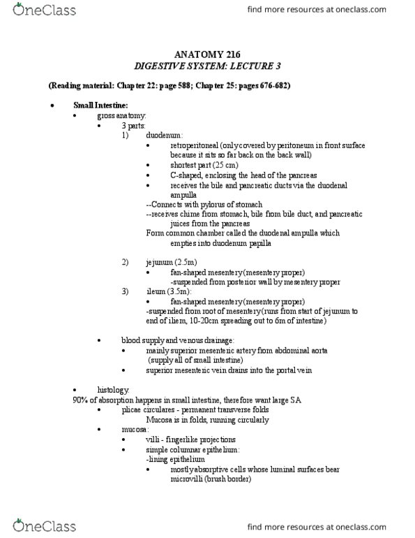
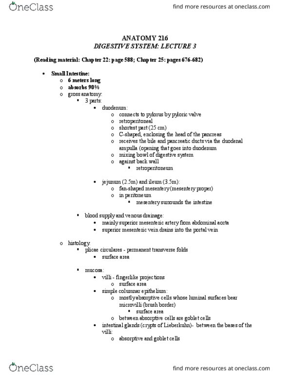
3 parts: duodenum: connects to pyloris of stomach separated by pyloric valve retroperitoneal: covered on front by peritoneum rest of bowel is suspended by mesentery shortest part (25 cm) Mostly absorptive cells whose luminal surfaces bear microvilli (brush border) facing lumen of gut between absorptive cells are goblet cells (mucus secreting) intestinal glands (crypts of lieberkuhn) - between the bases of the villi: absorptive and goblet cells. Acid starts eroding through he gut wall. Would have to get art of duodenum removed. Now they can treat ulcer with antibiotics so mucosa goes back to normal. Cecum, ascending colon, transverse colon, descending colon, sigmoid colon, rectum. Distinctive features: teniae coli, haustra, epiploic appendages ascending colon: retroperitoneal right colic (hepatic) flexure right angle turn just before liver transverse colon: transverse mesocolon: mesentery left colic (splenic) flexure another right angle turn. Descending colon: retroperitoneal becomes continuous with sigmoid colon sigmoid colon: sigmoid mesocolon: mesentery continues with rectum: rectum: