ANAT 216 Lecture Notes - Lecture 12: Common Hepatic Duct, Common Bile Duct, Hepatic Veins

28 Apr 2016
School
Department
Course
Professor
Document Summary
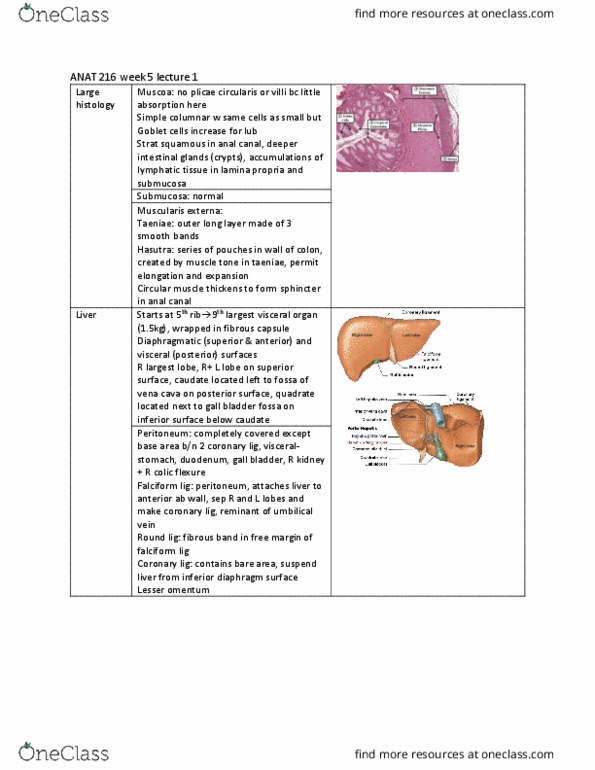
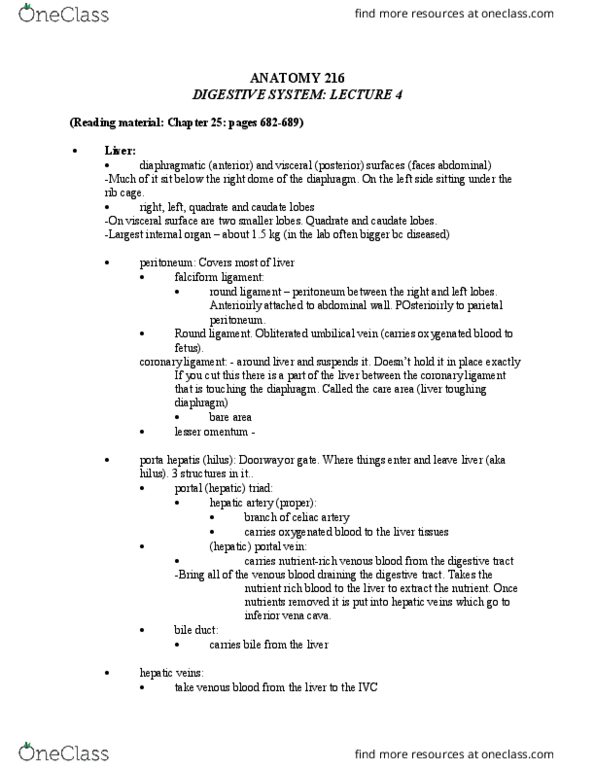
Liver: diaphragmatic (anterior): convex, visceral (posterior) surfaces: flat or concave, lobes of liver right, left, quadrate and caudate lobes. On right side but projects to the left. Under ribcage right lobe: bigger left lobe: smaller. Neck: goes into duct cystic duct spiral formation so it never closes stores and concentrates bile not immediately required for digestion squirted into duodenum. Billi rubin collects in blood and makes people ill skin gets yellow and whites of eye yellows: jaundice excess bile goes to gall bladder. Blood supply: cystic artery gallbladder stones tries to pass it if it goes into common duct very painful: histology: simple columnar epithelium with apical microvilli. Muscularis externa is scattered muscle fibers smooth muscle cells in muscularis. Pancreas: gross anatomy: retroperitoneal: lies behind stomach. Head lies in concavity of duodenum elongated body passes to left, becoming the tail which reaches spleen.