BPK 306 Lecture Notes - Lecture 26: Outer Plexiform Layer, Opsin, Outer Nuclear Layer

5 Dec 2017
School
Department
Course
Professor
Document Summary
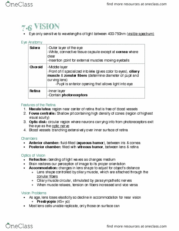
Basic anatomy: 3 concentric layers, outer fibrous coat (cornea) and epithelium (sclera, middle vascular coat (iris and choroid) Muscles: extraocular muscles innervated by cranial nerves iii, iv, and vi, dialator (sympathetic, sphincter (parasympathetic) Functions: detects and interprets electromagnetic waves between 400-750nm, brightness (intensity/number of photoreceptors) vs. wavelength (determines colour) Path of light: cornea -> aqueous humor -> lens -> vitreous humor -> focused on retina by lens and cornea. Iris = coloured portion: optic disc = blind spot, has no photoreceptors, choroid = structural and metabolic support. 2: fovea/macula = center of visual field, area of clearest vision -> higher density of photoreceptors than in the periphery. 1) retinal pigment epithelium (rpe: where phototransduction occurs (light to electrical signal, layer of pigment producing cells, melanin absorbs and diffracts light and prevents scatter of photons between photoreceptors (prs, provides nutrients, removes waste -> metabolic functions.