CRIM 356 Lecture Notes - Lecture 3: Confocal Microscopy, Sky Burial, Forensic Anthropology

29 Sep 2020
School
Department
Course
Professor
Document Summary
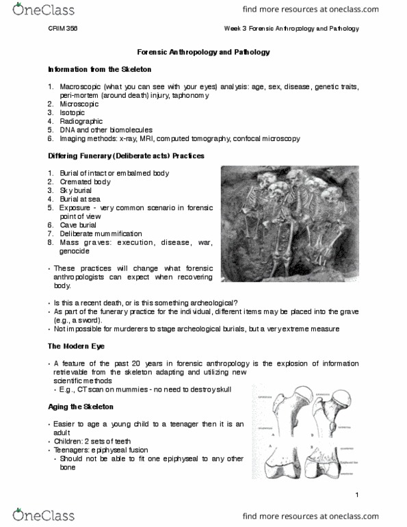
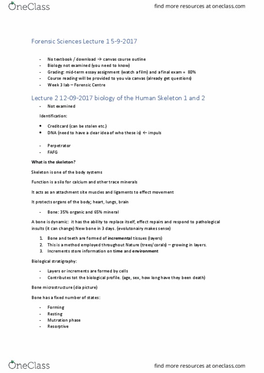
Crim 356 - lecture 3- forensic anthropology & pathology. Macroscopic analysis - things that can be seen. Imaging methods - x-ray, mri, computed tomography, confocal microscopy. Body cut up into pieces for birds to take away. Cannot tell from looking at body how old someone is. Feature of past 20 years in forensic anthropology - explosion of info retrievable from skeleton adapting & utilizing new scientific methodologies. Darker areas in x-ray - represent ossification of bones. Bones first begin to appear in third/fourth week of embryonic development. Clavicle bone - usually first to ossify. Light areas - indicate cartilage not yet replaced by bone. Epiphysis - fuses at diff points in development. Looking at this - key method of aging. Staged fusion at diff skeletal sites - indicative of individual"s age. Populations vary & so fusion is given as set of ranges. Important to note there are many more bones in childhood skeleton than the adult.