PSYC 3270 Lecture Notes - Lecture 4: Retinotopy, Radiography, Retina

27 Nov 2017
School
Department
Course
Professor
Document Summary
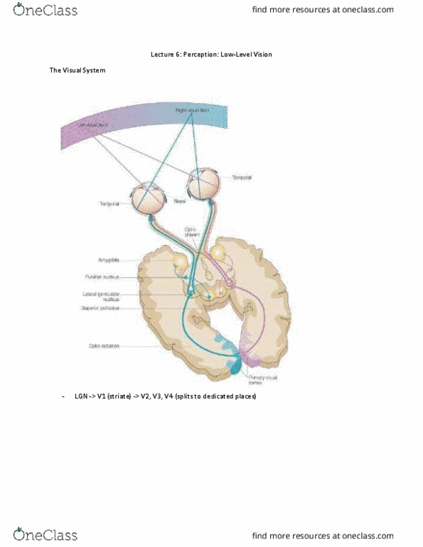
Understanding the organization of the brain and how that affect function. As light enters the eye, the object/image is inverted. Right side of object projects back to the left side of eyeball, vice versa contralateral. Distribution of photoreceptors on retina is not homogenous. Fovea: located in the center of the retina, photoreceptors are extremely dense and compact get more spatial info than other areas when you(cid:859)re looki(cid:374)g at so(cid:373)ethi(cid:374)g, your fovea is concentrated receive high detailed info. We see the same retinotopic organization even in v2,v3,v4. Cortical magnification: large representation of fovea (f) more cortical representation dedicated to center of the visual field. Inverted: superior (s) visual field projects to inferior (i) cortex and vice versa. Regions of v1 that were most radioactive (absorbed the most glucose) 1,2,3 = inner to outer rings (superior and inferior are flipped upside down, i. e. superior portions are presenting inferior parts of the image.