BIOL 1410 Lecture Notes - Lecture 7: Epithelium, Free Surface, Histology

1 May 2018
School
Department
Course
Professor

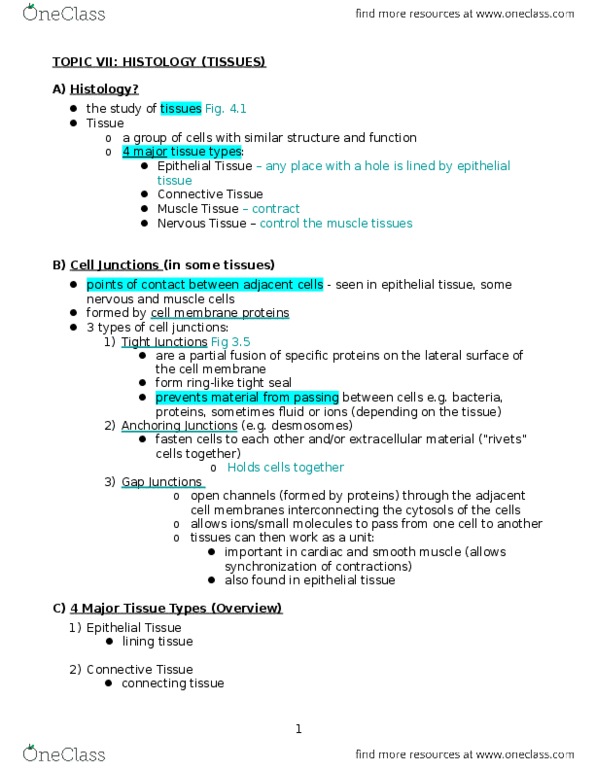
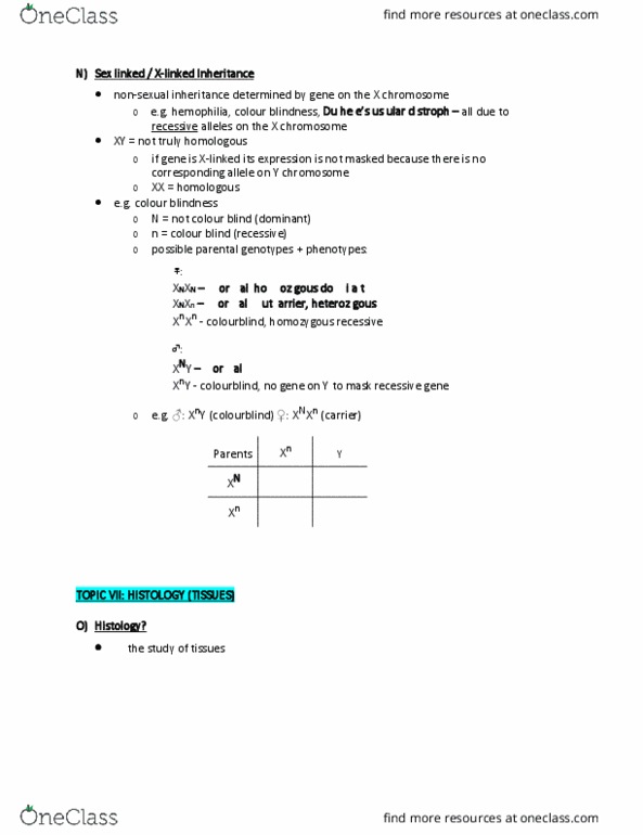
TOPIC 7: HISTOLOGY (TISSUES)
A) Histology?
the study of tissues
Tissue
o a group of cells with similar structure and function
o 4 major tissue types:
Epithelial Tissue
Connective Tissue
Muscle Tissue
Nervous Tissue
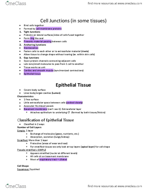
A) Cell Junctions (in some tissues) figure 4.3b
points of contact between adjacent cells - seen in epithelial tissue, some
nervous and muscle cells
formed by cell membrane proteins
3 types of cell junctions: anytime you have a junction, you’re making them up
from proteins
1) Tight Junctions figure 3.5
Holding the cells tightly together – prevents things from passing
through them
are a partial fusion of specific proteins on the lateral surface of
the cell membrane
form ring-like tight seal
prevents material from passing between cells e.g. bacteria,
proteins, sometimes fluid or ions (depending on the tissue)
2) Anchoring Junctions (e.g. desmosomes)
They are anchoring (holding) one cells to the next
proteins that fasten cells to each other and/or extracellular
material (“rivets” cells together – anchoring cells to cells)
3) Gap Junctions figure 3.5
open channels (formed by proteins) through the adjacent cell
membranes interconnecting the cytosols of the cells
allows ions/small molecules to pass from one cell to another
tissues can then work as a unit:
o important in cardiac and smooth muscle (allows
synchronization of contractions)
o also found in epithelial tissue
B) 4 Major Tissue Types (Overview)
1) Epithelial Tissue
lining tissue
find more resources at oneclass.com
find more resources at oneclass.com

2) Connective Tissue
connecting tissue
3) Muscle Tissue
contractile tissue
4) Nervous Tissue
signaling tissue
➢ Epithelial Tissue
covers body surface (skin)
lines body/organ cavities (from the outside following in)
o organ cavity = lumen – is a space/cavity not a tissue
Characteristics of Epithelia: “what are the overall characteristics of Epthelia?”
o has one free surface – does not have to be the ‘outside air’; it can be in
contact with the blood or surrounded by cell rings – its not = to air
o little extracellular space between cells
o avascular – no blood vessels
o Basement membrane – its bellow the epithelia tissue*
▪ extracellular layer
▪ attaches epithelium to underlying CT layer (formed by both
tissues – “velcro”)
Classification of Epithelia:
o most subtypes are classified + named according to:
▪ # of cell layers sitting on the basement membrane
• one layer = simple
• more than one layer = stratified
▪ shape of the cells in the apical layer (= layer touching the free
surface “so like the first row of cells”)
• flattened = squamous
• round or cube shaped = cuboidal
• rectangular = columnar
1) Simple Epithelia = 1 layer
▪ allow exchange of molecules (gasses, nutrients, etc.) –
absorption/secretion (getting things in or out)
▪ subtypes:
a) simple squamous (flattened) = 1 layer of squished (flat) cells
e.g. lungs
b) simple cuboidal (look like little cubes)= 1 layer of cube
shaped cells
e.g. kidneys
find more resources at oneclass.com
find more resources at oneclass.com
Document Summary
Tissue: a group of cells with similar structure and function, 4 major tissue types: Nervous tissue: cell junctions (in some tissues) figure 4. 3b. Points of contact between adjacent cells - seen in epithelial tissue, some nervous and muscle cells. 3 types of cell junctions: anytime you have a junction, you"re making them up from proteins: tight junctions figure 3. 5. Holding the cells tightly together prevents things from passing through them. Are a partial fusion of specific proteins on the lateral surface of the cell membrane. Prevents material from passing between cells e. g. bacteria, proteins, sometimes fluid or ions (depending on the tissue: anchoring junctions (e. g. desmosomes) They are anchoring (holding) one cells to the next. Proteins that fasten cells to each other and/or extracellular material ( rivets cells together anchoring cells to cells: gap junctions figure 3. 5. Open channels (formed by proteins) through the adjacent cell membranes interconnecting the cytosols of the cells.