BIOL 3542 Lecture Notes - Lecture 9: G Cell, Acinus, Pepsin

26 Jun 2018
School
Department
Course
Professor

Human Physiology II
Chapter 21: The Digestive System
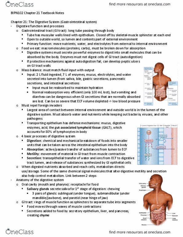
Anatomy of the Digestive System
digestive system begins with oral cavity (mouth, pharynx)
Gastrointestinal (GI) Tract: esophagus, stomach, small intestine, large intestine
long tube with muscular walls lined by secretory, transporting epithelium
rings of muscle (sphincters) separate tube into segments with distinct functions
food moves through tract propelled by waves of muscle contraction
Gut: portion of GI tract running from stomach to anus
products of digestion absorbed across intestinal epithelium, pass into interstitial fluid then
into blood or lymph for distribution throughout body
any waste remaining in lumen at end of GI tract leaves body through anus
because digestive system opens to outside, tract lumen and contents are actually part of
external environment
The Digestive System Is a Tube
1st stages of digestion begin with chewing, secretion of saliva by 3 pairs of salivary glands
sublingual glands under tongue
submandibular glands under mandible (jawbone)
parotid glands near hinge jaw
Esophagus: narrow tube that travels through thorax to abdomen
walls are skeletal muscle initially, transition to smooth muscle 2/3 way down length
ends at stomach just below diaphragm
Stomach: baglike organ that can hold up to 2L of food. fluid when fully expanded
continues digestion that began in mouth with acids, enzyme to create chime
Pylorus: opening between stomach, small intestine
Pyloric Valve: thickened band of smooth muscle that guards pylorus, relaxes to allow only small
amounts of chyme into the small intestine at a time
stomach is intermediary between behavioural act of eating, physiological events of digestion,
absorption in intestine
integrated signals, feedback loops between intestine, stomach regulate rate at which chyme
enters duodenum, ensuring intestine not overwhelmed with more than it can digest, absorb
3 parts of small intestine are duodenum, jejunum, ileum
digestion carried out by intestinal enzymes, aided by exocrine secretions from pancreas, liver
that enter initial section of duodenum through ducts
sphincter of Oddi keeps pancreatic fluid, bile from entering small intestine except during
meal
in colon, watery chyme becomes semisolid feces as water, electrolytes absorbed out, into
ECF
when feces propelled into terminal section of large intestine (rectum), distension of rectal
wall triggers defecation reflex
feces leave GI tract through anus

the external anal sphincter of skeletal muscle is under voluntary control
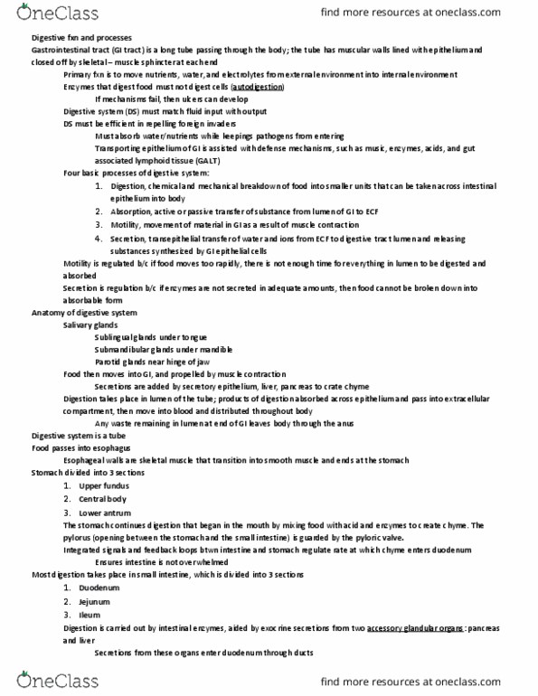
The GI Tract Has Four Layers
additional surface area added by tubular invagination of surface that extend down into
supporting connective tissue called gastric glands in stomach, crypts in intestine
deepest invaginations form secretory submucosal glands that open into lumen through ducts
gut wall consists of 4 layers:
1. inner mucosa facing lumen
2. submucosa
3. layers of smooth muscle known as muscularis externa
4. a covering of connective tissue called the serosa
Mucosa
Mucosa: inner lining of GI tract
3 layers:
1. single layer of mucosal epithelium facing lumen
cells of mucosa include transporting epithelial cells (called enterocytes in small
intestine), endocrine and exocrine secretory cells, and stem cells
at mucosal (apical) surface, cells secrete ions, enzymes, mucus, paracrine molecules
into lumen
on serosal (basolateral) surface, substances being absorbed from lumen, molecules
secreted by epithelial cells enter ECF
in stomach, colon, cell-to-cell junction form tight barrier so little can pass between
cells
intestinal epithelium considered “leaky” because some water, solutes can be absorbed
between cells (paracellular pathway) instead of through them; selectivity can be
regulated to some extent
GI stem cells are rapidly dividing, undifferentiated cells that continuously produce
new epithelium in crypts, gastric glands
as they divide, newly formed cells are pushed toward luminal surface of epithelium
rapid turnover, cell division rate in GI tract makes these organs susceptible to
developing cancers
2. Lamina Propria: sub-epithelial connective tissue that holds epithelium in place
contains nerve fibers, small blood and lymph vessels
absorbed nutrients pass into blood, lymph here
also contains wandering immune cells (macrophages, lymphocytes) patrolling for
invaders that enter through breaks in epithelium
in intestine, collections of lymphoid tissue adjoining the epithelium form small
nodules, larger Peyer’s patches that create visible bumps in mucosa
lymphoid aggregation major part of gut-associated lymphoid tissue (GALT)
3. Muscularis Mucosae: thin layer of smooth muscle
separates lamina propria from submucosa
contractions of muscles in this layer alter effective surface area for absorption by
moving villi back and forth

Submucosa
Submucosa: middle layer of gut wall composed of connective tissue with larger blood, lymph
vessels running through it
contains submucosal plexus which innervates cells in epithelial layer, smooth muscle of
muscularis mucosae
Muscularis Externa
outer wall of GI tract
consists primarily of 2 layers of smooth muscle: an inner circular layer and an outer
longitudinal layer
contraction of circular layer decreases diameter of lumen
contraction of longitudinal layer shortens tube
stomach has incomplete 3rd layer of oblique muscle between circular muscles, submucosa
Myenteric Plexus: 2nd nerve network of enteric nervous system, lies between longitudinal,
circular muscle layers and controls, coordinates motor activity of the muscularis externa
Serosa
Serosa: a connective tissue membrane that is continuation of the peritoneal membrane lining
the abdominal cavity
outer covering of entire digestive tract
peritoneum forms sheets of mesentery that hold intestines in place so they don’t become
tangled as they move
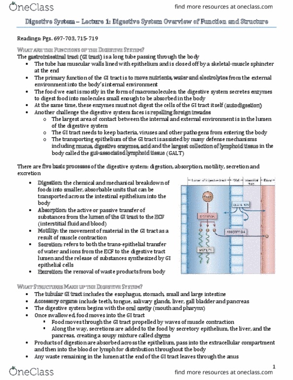
Digestive Function and Processes
primary function of digestive system to move nutrients, water, electrolytes from external
environment to body’s internal environment
Digestion: chemical, mechanical breakdown of foods into smaller units that can be taken across
intestinal epithelium into the body
Absorption: movement of substances from lumen of GI tract to ECF
Secretion: either movement of water, ions from ECF to digestive tract lumen (opposite of
absorption) OR release of substances synthesized by GI epithelial cells into lumen or ECF
Motility: movement of material in GI tract as result of muscle contraction
digestive system faces 3 challenges:
1. Avoiding autodigestion
enzymes mustn’t digest cells of GI tract itself
if protective mechanisms against autodigestion fail, raw patches called peptic ulcers
develop in walls of GI tract
2. Mass balance
maintaining mass balance by matching fluid input with output
normally intestinal reabsorption very efficient
vomiting, diarrhea can become emergencies when GI secretion lost to environment
instead of being reabsorbed
in severe cases, fluid loss can deplete ECF volume to point that circulatory system is
unable to maintain adequate blood pressure
3. Defense
Document Summary
Anatomy of the digestive system digestive system begins with oral cavity (mouth, pharynx) Gastrointestinal (gi) tract: esophagus, stomach, small intestine, large intestine. Long tube with muscular walls lined by secretory, transporting epithelium. Rings of muscle (sphincters) separate tube into segments with distinct functions. Food moves through tract propelled by waves of muscle contraction. 1st stages of digestion begin with chewing, secretion of saliva by 3 pairs of salivary glands. Esophagus: narrow tube that travels through thorax to abdomen. Walls are skeletal muscle initially, transition to smooth muscle 2/3 way down length. Stomach: baglike organ that can hold up to 2l of food. fluid when fully expanded. Continues digestion that began in mouth with acids, enzyme to create chime. When feces propelled into terminal section of large intestine (rectum), distension of rectal wall triggers defecation reflex feces leave gi tract through anus. The external anal sphincter of skeletal muscle is under voluntary control.