BIOL 3542 Lecture Notes - Lecture 17: Pericardium, Rib Cage, Breathing

26 Jun 2018
School
Department
Course
Professor

Human Physiology II
Chapter 17: Mechanics of Breathing
The Respiratory System
Cellular Respiration: intracellular reaction of oxygen with organic molecules to produce carbon
dioxide
External Respiration: movement of gases between environment and body cells
4 integrated processes:
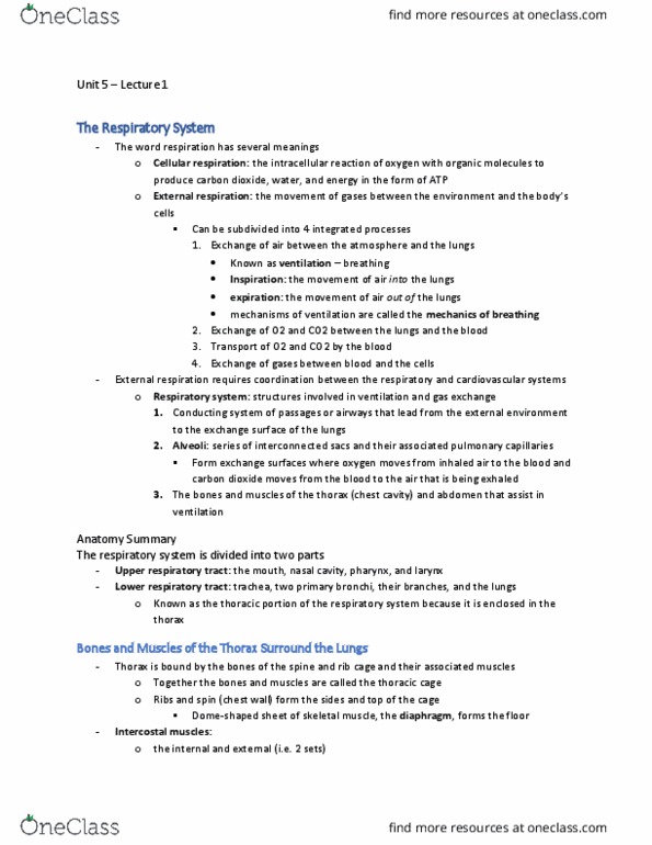
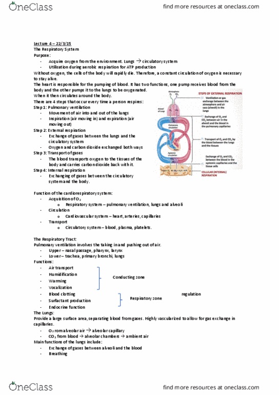
1. Ventilation: exchange of air between atmosphere and lungs
oInspiration: movement of air into lungs (inhalation)
oExpiration: movement of air out of lungs (exhalation)
oMechanisms of Breathing: mechanisms by which ventilation takes place
2. exchange of oxygen, carbon dioxide between lungs, blood
3. transport of oxygen, carbon dioxide by blood
4. exchange of gases between blood, cells
Respiratory System: structures involved in ventilation, gas exchange
1. Conducting System: airways that lead from external environment to exchange surface of
lungs
2. Alveoli: series of interconnected sacs, associated pulmonary capillaries that form
exchange surface where oxygen moves from inhaled air to blood, carbon dioxide from
blood to to-be-exhaled air
3. bones, muscles of thorax and abdomen that all assist in ventilation
Upper Respiratory Tract: mouth, nasal cavity, pharynx, larynx
Lower Respiratory Tract: trachea, 2 primary bronchi and their branches, lungs
a.k.a. thoracic portion of respiratory system because it’s enclosed in the thorax
Bones and Muscles of the Thorax Surround the Lungs
thorax bounded by bones of spine, rib cage, their associated muscles
Thoracic Cage: bones, muscles together
2 sets of intercostal muscles, internal and external, connect 12 pairs of ribs
sternocleidomastoid, scalene muscles run from head, neck to 1st 2 ribs
pericardial sac contains heart
pleural sacs each surround a lung
esophagus, thoracic blood vessels, nerves pass between pleural sacs
Pleural Sacs Enclose the Lungs
Lungs: light, spongy tissue, volume occupied by air-filled spaces
cone-shaped
nearly fill thoracic cavity
bases rest on diaphragm
bronchi connect lungs to trachea
each lung surrounded by double-walled pleural sac, membranes line inside of thorax, cover
outside of lungs
find more resources at oneclass.com
find more resources at oneclass.com

pleura contain several layers of elastic connective tissue, numerous capillaries
opposing layers held together by thin film of pleural fluid
pleural fluid creates moist surface so opposing membranes can slide across one another as
lungs move, holds lungs tight against thoracic wall
Airways Connect Lungs to the External Environment
air flows from pharynx, through larynx to trachea
larynx contains vocal cords, connective tissue bands that vibrate, tighten to create sound
when air moves past
Trachea: semi-flexible tube held open by C-shaped cartilage rings, extending down into thorax
branches in pair of primary bronchi, 1 in each lung
Bronchi: semi-rigid tubes supported by cartilage
branch repeatedly in lungs into progressively smaller bronchi (bronchioles)
Bronchioles: small, collapsible passageways with walls of smooth muscle
continue branching until respiratory bronchioles form transition between airways,
exchange epithelium of lung
total cross-sectional area increases with each division of airways
lowest in upper respiratory tract, greatest in bronchioles
velocity of air flow inversely proportional to total cross-sectional area of airways
greatest in upper airways, slowest in terminal bronchioles
The Airways Warm, Humidify, and Filter Inspired Air
upper airways, bronchi condition air before it reaches alveoli
3 components of conditioning:
1. warming air to body temp. (37oC) so core body temp. doesn’t change, alveoli aren’t
damaged by cold air
2. adding water vapour until air reaches 100% humidity, so moist exchange epithelium
doesn’t dry out
3. filtering out foreign material so viruses, bacteria, inorganic particles don’t reach alveoli
breathing through mouth not as effective at warming, moistening air as breathing through
nose
air filtered in trachea, bronchi lined with ciliated epithelium whose cilia bathed in watery
saline layer produced by epithelial cells with chlorine ions secreted into lumen by apical
anion channels draw sodium ions into lumen through paracellular pathway
movement of solute from ECF to lumen creates osmotic gradient, water follows ions into
airways
CTFR channel one ion channel found in apical surface of epithelium
sticky mucus layer secreted by goblet cells in epithelium floats over cilia to trap inhaled
particles
contains immunoglobulins that disable many pathogens
Mucociliary Escalator: cilia beat in upward motion that moves mucus towards pharynx
once reaching pharynx, mucus can be spit out (expectorated) or swallowed
stomach acid, enzymes destroy remaining microorganisms
in cystic fibrosis, inadequate ion secretion decreases fluid movement in airways
find more resources at oneclass.com
find more resources at oneclass.com

without saline layer, cilia become trapped in thick, sticky mucus, cannot move
mucus cannot be cleared, bacteria colonize airways resulting in recurrent lung infections
Alveoli Are the Site of Gas Exchange
air-filled alveoli, clustered at ends of terminal bronchioles, make up bulk of lung tissue
primary function is gas exchange between themselves, blood
each alveolus composed of single layer of epithelium
type I alveolar cells, larger cells used for gas exchange
layer of basement membrane fuses alveolar epithelium to capillary endothelium
remaining area filled with small amount of interstitial fluid
smaller, thicker type II alveolar cells synthesize, secrete chemical surfactant that mixes
with thin fluid lining of alveoli to aid lungs as they expand during breathing
minimize amount of fluid present in alveoli by transporting solutes, followed by water,
out of alveolar air space
thin walls of alveoli don’t contain muscle, would block rapid gas exchange, so lung itself
cannot contract
connective tissue between alveolar epithelial cells contains elastin, collagen fibers that
create elastic recoil when lung tissue stretched
blood vessels fill space between alveoli, forming “sheet” of blood in close contact with air-
filled alveoli, allowing for rapid gas exchange
Pulmonary Circulation Is High-Flow, Low Pressure
pulmonary trunk gets low-oxygen blood from right ventricle, divides into 2 pulmonary
arteries, 1 to each lung
oxygenated blood from lungs returns to left atrium via pulmonary veins
rate of blood flow through lungs much higher than in other tissues
as much blood flows through lungs in 1min than through rest of body in same amount of
time
pulmonary blood pressure low
right ventricle doesn’t have to pump as forcefully to create blood flow through lungs because
resistance of pulmonary circulation low
attributed to shorter total length of pulmonary blood vessels, distensibility, large total
cross-sectional area of pulmonary arterioles
normally net hydrostatic pressure filtering fluid out of a pulmonary capillary into interstitial
space low because of low mean blood pressure
lymphatic system removes filtered fluid, lung interstitial fluid volume minimal, resulting in
short distance between alveolar air space, capillary endothelium, allowing rapid diffusion of
gases between them
Gas Laws
Air Is a Mixture of Gases
Dalton’s Law: total pressure exerted by mixture of gases is sum of pressures exerted by
individual gases
Partial Pressure (Pgas): pressure of single gas in mixture
find more resources at oneclass.com
find more resources at oneclass.com
Document Summary
Cellular respiration: intracellular reaction of oxygen with organic molecules to produce carbon dioxide. External respiration: movement of gases between environment and body cells. 3. transport of oxygen, carbon dioxide by blood: exchange of gases between blood, cells. Upper respiratory tract: mouth, nasal cavity, pharynx, larynx. Lower respiratory tract: trachea, 2 primary bronchi and their branches, lungs. A. k. a. thoracic portion of respiratory system because it"s enclosed in the thorax. Bones and muscles of the thorax surround the lungs thorax bounded by bones of spine, rib cage, their associated muscles. 2 sets of intercostal muscles, internal and external, connect 12 pairs of ribs sternocleidomastoid, scalene muscles run from head, neck to 1st 2 ribs pericardial sac contains heart. Pleural sacs each surround a lung esophagus, thoracic blood vessels, nerves pass between pleural sacs. Lungs: light, spongy tissue, volume occupied by air-filled spaces.