ANP 1105 Lecture Notes - Lecture 12: Superior Vena Cava, Pulmonary Valve, Coronary Sinus

19 Jan 2016
School
Department
Course
Professor
Document Summary
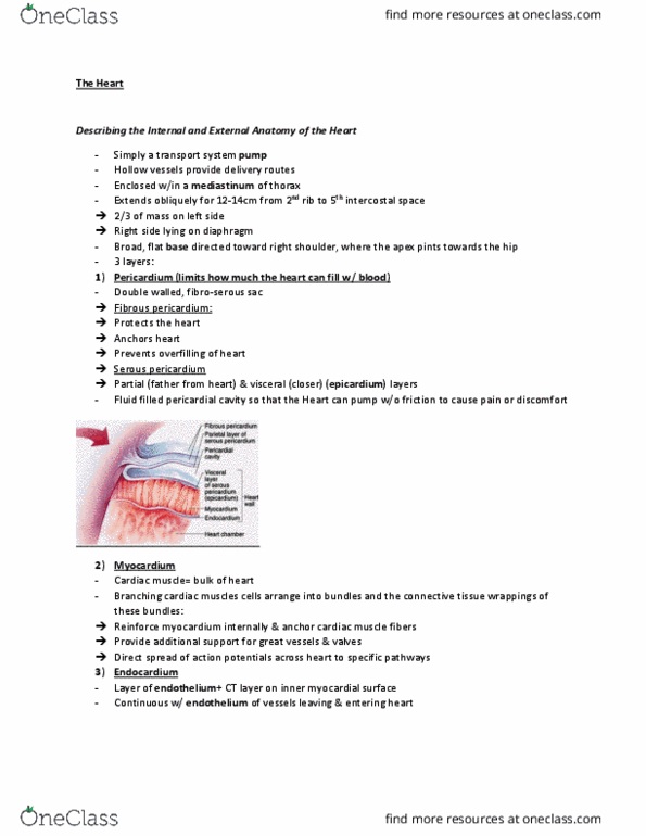
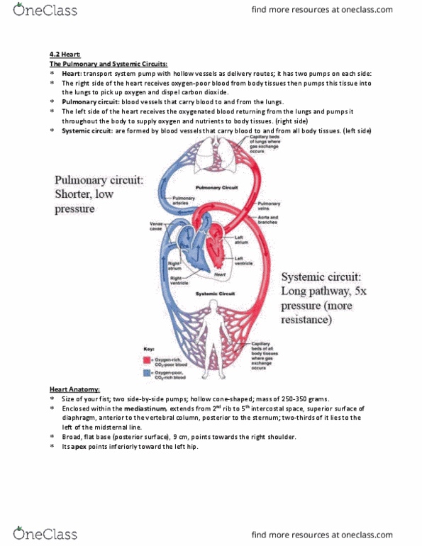
A transport system pump and the hollow blood vessels provide delivery routes. Fibrous pericardium: protects heart, anchors heart, prevents overfilling of heart. Serous pericardium: parietal and visceral epicardium layers. Provide additional support for great vessels and valves. Direct spread of action potentials across hear to specific pathways. Continuous with endothelium of vessels leaving and entering the heart. Interatrial septa wall in between the two atria. Small, thin walled and only need to convey blood to ventricles. Deoxygenated blood enters right atrium via superior vena cava, inferior vena cava, and coronary sinus. Oxygenated blood returns from the left atrium from the four pulmonary veins. Pumps of the heart, walls are thick. Right ventricle pumps bloods to the pulmonary trunk. Internal walls have muscle bundles; trabeculae carneae (rounded or irregular muscular columns that project from the inner surface of the right and left ventricles of the heart), papillary muscles (valve function)