ANP 1106 Lecture Notes - Lecture 3: Electrochemical Gradient, Tubuloglomerular Feedback, Vasoconstriction

6 Jun 2018
School
Department
Course
Professor

Anatomy and Physiology 1107
Notes: Lecture 12
Prof: J. Carnegie
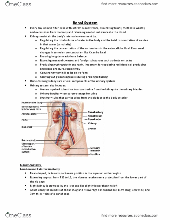
➢ The renal system
o It is bean shaped and retroperitoneal
o Superior lumbar region: from the 12th thoracic vertebra to the 3rd lumbar
vertebra
o It has some protection from the rib cage, the kidneys are pushed lower by the
liver
o The adrenal glands sit on top of the kidneys
➢ There are three layers of supportive tissue
▪ Real apsule: it is firous, adheres diretl to the kide’s surfae ad is
a strong barrier to ____
▪ Perirenal fat capsule: it cushions and helps holds the kidneys in place
• Renal ptosis: it is a blockage of urine flow due to a kink in the
urethers which causes the urine to go back into the kidneys
▪ Renal fascia: dense CT surrounds the adrenal glands and the kidney, it
plays an anchoring role
➢ Internal anatomy
o Cortex: where the actual filtration of the blood occurs
o Medulla: also called medullary/renal pyramids, it is a darker colour and appears
striped because they represent tiny collection ducts that carry filtered urine to
the ureters, it is separated by renal columns, each medullary unit = 1/8 of the
kidney
o Pelvis: it is a flat, funnel-shaped tube that is continuous with the ureter, major
and minor calices enclose the papillae of the pyramids and calices collect urine,
walls of calyces, the pelvis and the ureter contain smooth muscle and propel the
urine by peristalsis (Two or three minor calices lead into a major calice which
then leads to a single unit of the pelvis which leads into the ureter)
o Pyelitis is an infection that has reached the tubing (ureter and pelvis)
o Pyelonephritis is an infection that has reached the kidneys
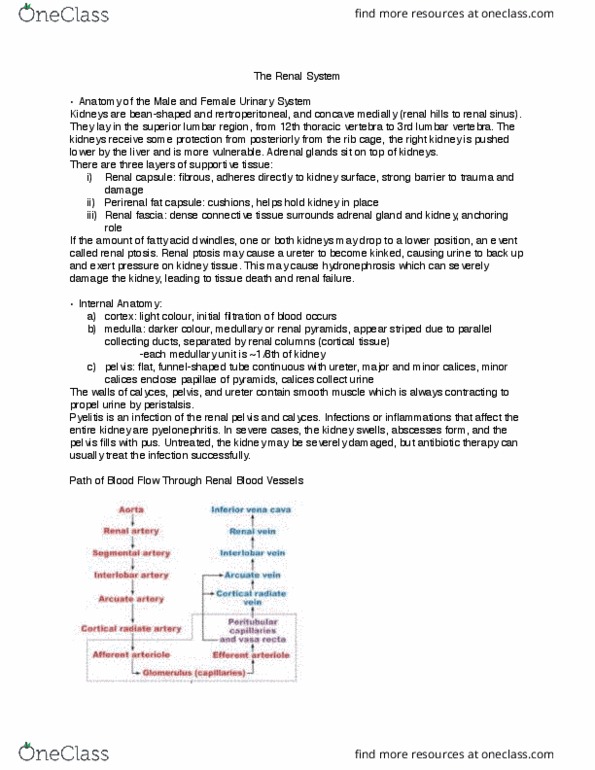
➢ Blood and nerve supply
o Renal arteries: ¼ total systemic cardiac output to the kidneys per minute
o Arterial branches pass up between medullary pyramids to reach the cortex,
venous branches drain back via the same route
o The nerve supply provided by the renal plexus of primarily sympathetic fibers
regulate the renal blood flow by adjusting the diameters of renal arterioles
➢ Nephrons
o There are 106 nephrons and thousands of collecting ducts in each kidney
find more resources at oneclass.com
find more resources at oneclass.com

▪ Gloerulus, Boa’s apsule, real apsule, feestrated gloerular
endothelium, podocytes (cells in the inner layer), pedicles (projections
that stick out of podocytes) and filtration slits
o Proximal convoluted tubule → loop of Henle → distal convoluted tubule →
collecting duct → papillary duct → minor calyx
▪ The collecting duct has
• Priipal ells (laks/sparse iroilli ad aitais the od’s
salt and water balance)
• Intercalated cells (abundant microvilli and maintains the acid-base
balance of the body)
o There are two types of nephrons
▪ Cortical (85% of nephrons)
• Useful for all functions
▪ Juxtamedullary (15% of nephrons)
• Useful for conserving fluids, it is important during dehydration
o Microcirculation of the nephron
• Glomerulus: it is specialized for filtration and both fed and drained
by the arterioles
o Arterioles are high resistance vessels
o The afferent arteriole has a larger diameter because more
blood goes in than comes out
• Peritubular Capillaries: arise from efferent arterioles and drain
into renal venules
• Vasa Recta: helps conserve water by moving it into the blood
stream
o They are found paralleling the longest nephron loops
➢ Juxtaglomerular apparatus
o It is at the junction of the early distal convoluted tubule and afferent/efferent
arterioles, it regulates renal function
▪ Arteriole walls (Juxtaglomerular cells/granular cells)
• They are enlarged smooth muscle cells that secrete renin, they act
as mechanoreceptors (they are stretch receptors that determine
blood pressure)
▪ Tubule wall (macula densa cells)
• They are chemo/osmo-receptors that monitor the filtrate and
adjust the glomerular filtration rate (it monitors the osmolarity to
determine how concentrated the filtrate is in order to determine
how fast it filters)
o It regulates filtrate formation and systemic blood pressure
➢ Micturition pathway
o It is also called urination; it is the act of emptying the urinary bladder
find more resources at oneclass.com
find more resources at oneclass.com

▪ The bladder expands to accumulate urine, it is lined by transitional
epithelium (stretch epithelium)
o Trigone
▪ It is composed of smooth muscle, when you empty your bladder your
urine will sit there. It is important clinically because it is most likely where
you will get an infection
o Internal urethral sphincter
▪ It is smooth muscle that is involuntarily controlled
o External urethral sphincter
▪ It is skeletal muscles that is voluntarily controlled
➢ Renal Physiology
o They are major excretory organs and are a perfect examples of homeostatic
organs
o They major function is to filter several litres of fluid from the bloodstream daily
▪ Toxins, metabolic wastes and excess ions leave the body in the urine
▪ Materials that are still needed by the body are returned in the
bloodstream
o Additional functions
▪ Regulate the blood and volume composition (ex: blood/water and
acid/base)
▪ Produce the enzyme renin to help regulate blood pressure and kidney
function
▪ Produce the hormone erythropoietin to stimulate the production of RBCs
▪ Metabolize vitamin D to its active form
o 1000-1200ml of blood passes through the glomeruli each minute (approximately
650ml is plasma, of this 120-125ml of plasma is forced into the renal tubules
every minute), this is the equivalence to filtering the entire plasma volume over
60 times/day
o Filtrate = plasma minus the proteins
o Urine = filtrate minus the nutrients, essential ions and water
o The kidneys process approximately 180L of fluid/day and only 1% of this is urine
▪ Glomerular filtration
• It is a passive process in which hydrostatic pressure forces fluids
and solutes through a membrane
• It is very efficient because
o The filtration membrane is 1000s times more permeable
than other capillary membranes
o The glomerular blood pressure is higher than in other
capillary beds (55mm Hg vs < 18mm Hg), blood pressure is
able to be maintained at such a high level by regulating the
diameters/constantly adjusting the vessels
o 180L of filtrate formed by kidney capillaries vs 3-4L by all
other capillary beds combined
find more resources at oneclass.com
find more resources at oneclass.com
Document Summary
It is bean shaped and retroperitoneal: superior lumbar region: from the 12th thoracic vertebra to the 3rd lumbar vertebra. It has some protection from the rib cage, the kidneys are pushed lower by the liver: the adrenal glands sit on top of the kidneys. Intercalated cells (abundant microvilli and maintains the acid-base balance of the body: there are two types of nephrons, cortical (85% of nephrons, useful for all functions. It regulates filtrate formation and systemic blood pressure. It is also called urination; it is the act of emptying the urinary bladder: the bladder expands to accumulate urine, it is lined by transitional epithelium (stretch epithelium, trigone. It is composed of smooth muscle, when you empty your bladder your urine will sit there. It is important clinically because it is most likely where you will get an infection. It is smooth muscle that is involuntarily controlled: external urethral sphincter. It is skeletal muscles that is voluntarily controlled.