BCH 4101 Lecture Notes - Lecture 2: Histone H3, Cenpa, Sister Chromatids

28 Apr 2018
School
Department
Course
Professor

September 13, 2017
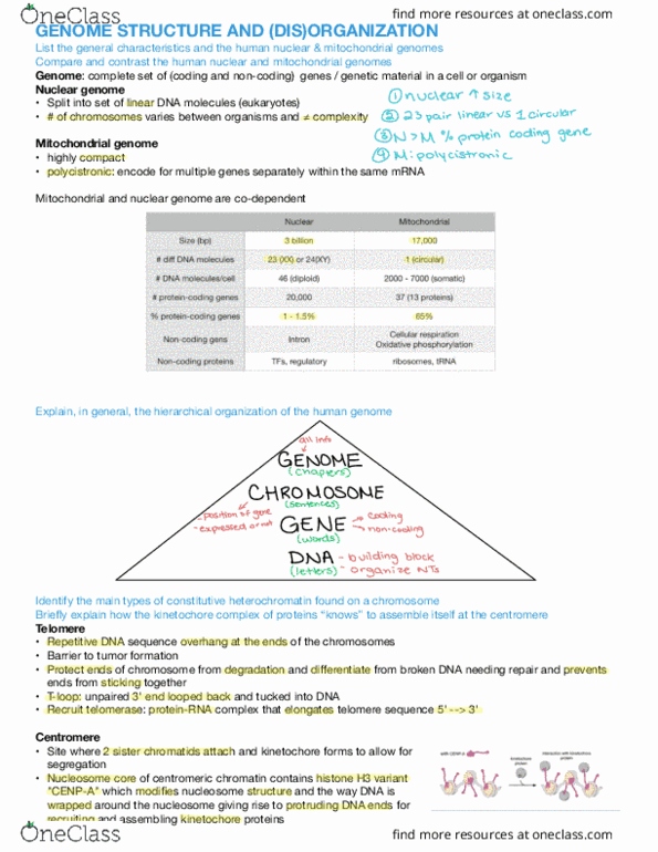
Genome (Dis)organization
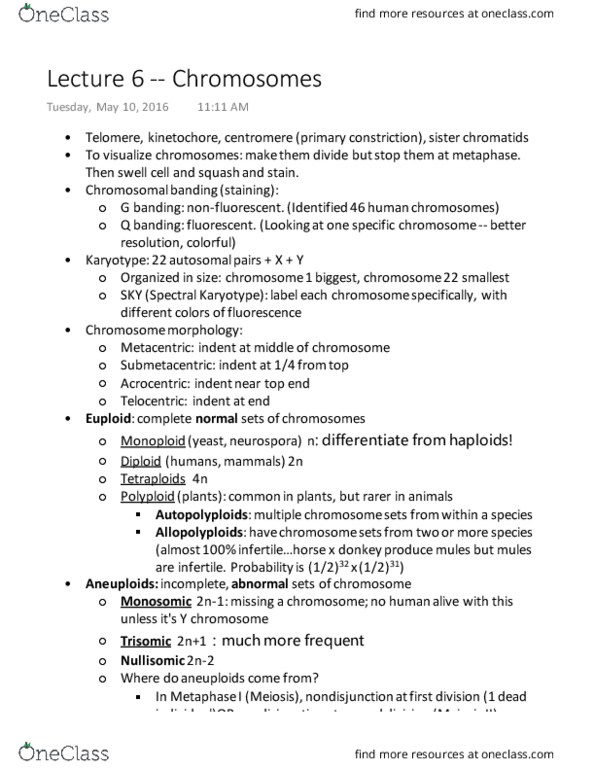
Chromosome Structure
Consists of two sister chromatids connected at the centromere
The telomere:
-Repetitive DNA sequence (5’-TTAGGG-3’) overhang at the ends of the chromosome
-Protects the integrity of the genetic material
-The very end of the sequence can’t be copied
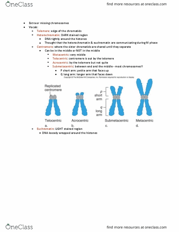
The centromere: interacts with kinetochore proteins (required for mitosis)
-Centromeric chromatin contains CENP-A (histone H3 variant), which attracts the proteins that form the kinetochore
-Allows for proper kinetochore assembly at the correct location
-Centromere positions varies and are characteristic for each chromosome
•Metacentric = in the middle
•Submetacentric = close to the middle
•Acrocentric = close to the end
•Telocentric = at the end (at the telomere)
Studying Chromosomes
Karyotype: an individual’s chromosome collection
-The characteristics of someone’s chromosomes
-Ex. Number, size, shape
-Ambiguous identification: chromosomes can look very similar under the microscope
-Human genome is separated into 7 groups, based on relative size and position of the centromere
•Largest chromosome = chromosome 1, smallest chromosome = chromosome 22, sex chromosomes are always
put last
Human Karyotyping
Karyogram: photo of an individual’s chromosomes, sorted and arranged by size
-Photo of someone’s karyotype
Karyotyping: creating a karyogram
1

September 13, 2017
Cytogenetic staining techniques for chromosome banding patterns: highlight the relative pattern of euchromatin and
heterochromatin in order to better distinguish between chromosomes
-Each chromosome will have a unique patten of bands when we apply a particular stain
-G-banding: stained with Giemsa
•Most common
•Dark bands (G-bands) are AT-rich: represents heterochromatin
-Stain very darkly
•Light bands are GC-rich: represents euchromatin
-Stain very faintly
•Metaphase chromosomes must be used: the point where the chromosomes are in their most condensed state,
making the banding pattern of compact vs. less compact DNA more easily observable
•Pre-treatment consists of mild proteolysis, making the chromosomes more susceptible to being bound by the
dye
-R-banding: also stained with Giemsa but differs in the identity of the G-bands and the pre-treatment
•Dark bands (G-bands) are GC-rich: represents euchromatin
•Light bands are AT-rich: represents heterochromatin
-AT-rich regions are more susceptible to denaturation, so they appear lighter
•Metaphase chromosomes
•Pre-treatment consists of heat denaturation
•Less commonly used than G-banding
-Q-banding: stained with quinacrine
•Fluorescent stain
•Binds both AT- and GC-rich regions, but only AT-quinacrine complexes fluoresces
-Only heterochromatin fluoresces
•Preferentially stains heterochromatin
•Uses metaphase chromosomes
•Useful for studying Y-chromosome
•No pretreatment required
-C-banding: stained with Giemsa, differs with the pretreatment
•Stains constitutive heterochromatin - stains the centromere
•Uses metaphase chromosomes
2

September 13, 2017
•Alkaline denaturation prior to staining
Idiogram: reference map showing expected banding patterns for each chromosome
-Used as a reference to order chromosomes into a karyogram
-International System for Cytogenetic Nomenclature (ISCN): standardized way of referring to
chromosomal conditions
•Everything is named in reference to the centromere - everything is numbered in reference to the
centromere
•Short arm (p - petite): always oriented towards the top
•Long arm (q - queue = long line): always oriented towards the bottom
•Numerical position: numbered outwards on each arm started from the centromere, larger number
= farther from centromere
-Each numerical block is subdivided into regions
-Sometimes there is a period followed by another number, representing subbannds
-Ex. 4q26.2: represents chromosome 4, q arm, region 2, band 6 within region 2, subband 2
Karyotype and Disease
Analysis of band patterns can be used to investigate chromosomes abnormalities (visible alterations)
-Changes need to be large enough to be seen
-Ex. Point mutations can’t be visualized but gross chromosomal abnormalities (deletions, extra chromosomes, etc.)
can be
2 types of abnormalities:
-Constitutional (familial) abnormality: inherited
•Present in all cells of the body - come from the germ cell
•NB: mosaic expression is possible in familial abnormality, but this is not usually the case
-Somatic abnormality: acquired
•Can be acquired in response to mutagens
•Mosaic expression: some cells will have it and some cells won’t
Abnormalities can be further divided into numerical or structural abnormalities
Numerical Abnormalities
Refers to aneuploidy: gain or loss of complete chromosomes
-Abnormal number of chromosomes
3
Document Summary
Consists of two sister chromatids connected at the centromere. Repetitive dna sequence (5"-ttaggg-3") overhang at the ends of the chromosome. Protects the integrity of the genetic material. The very end of the sequence can"t be copied. The centromere: interacts with kinetochore proteins (required for mitosis) Centromeric chromatin contains cenp-a (histone h3 variant), which attracts the proteins that form the kinetochore. Allows for proper kinetochore assembly at the correct location. Centromere positions varies and are characteristic for each chromosome: metacentric = in the middle, submetacentric = close to the middle, acrocentric = close to the end, telocentric = at the end (at the telomere) Ambiguous identi cation: chromosomes can look very similar under the microscope. Human genome is separated into 7 groups, based on relative size and position of the centromere: largest chromosome = chromosome 1, smallest chromosome = chromosome 22, sex chromosomes are always put last. Karyogram: photo of an individual"s chromosomes, sorted and arranged by size.