PHS 3342 Lecture Notes - Lecture 6: Pulmonary Circulation, Pulmonary Vein, Mediastinum

28 Apr 2018
School
Department
Course
Professor

January 23, 2018
Cardiac Physiology
Overview of the Cardiovascular System
Role: bring oxygen and nutrients to tissues, remove wastes
Heart is simply 2 pumps, hollow blood vessels are delivery tubes
-Arteries: carry blood away from the heart
-Veins: carry blood to the heart
Each beat of the heart (cardiac cycle) requires the coordination of many events
2 circuits:
-Pulmonary circulation: brings blood from the heart to the lungs for gas exchange
•Has serial blood flow - sequential changes to all of the blood following the pathway (ex. Addition of O2, removal
of CO2)
-Systemic circulation: distributes oxygen-rich blood to the body, returns oxygen-deprived blood to the heart
•Has parallel blood flow - blood subdivided to service many vascular beds at one
•Relative distributions can be adjusted as per body’s need
Equal volumes flowing in pulmonary and systemic circuits, but the two ventricles have unequal workloads
-Pulmonary circuit (right ventricle): short, low pressure circulation
•Distance is short, so there is little resistance
-Systemic circuit (left ventricle): long pathways with 5x the resistance
•Much longer pathway, so there is a greater cumulative resistance to flow - left ventricle needs to pump much
harder in order to ensure that the blood can make it through the entire pathway
•Walls of the left ventricle are ~3x as thick as those of the right ventricle
The Heart: a Muscular Pump
Located in the mediastinum of the thorax
12-14 cm in length, 250-350 mg
-*Don’t need to memorize numbers
Apex points to the left, oblique position in thorax (⅔ left of midsternum)
1

January 23, 2018
Walls of the Heart
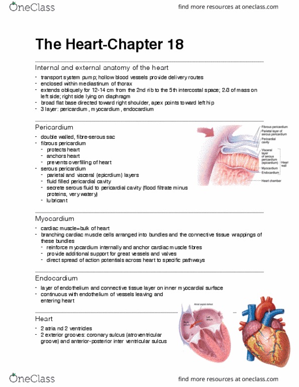
3 layers:
-Pericardium: double-walled, fibroserous
•Fibrous pericardium: tough, outermost layer of CT
-Protects the heart - protects against bacterial infection
-Anchors heart to diaphragm, great vessels
-Prevents overfilling of heart
•Serous pericardium (aka epicardium): parietal and visceral layers
separated by fluid-filled pericardial cavity
-Parietal: outer layer
-Visceral layer: inner layer
-Fluid allows the heart to beat without causing discomfort - decreases
friction
-Myocardium: cardiac muscle
•Branching cardiac muscle cells arranged in spiral/circular bundles
-Endocardium: endothelium + CT layer on the inner myocardial surface
•Continuous with endothelium of vessels entering and leaving the heart
CT:
-Provides strength for network
-Support for great vessels, valves
-Directs spread of AP across heart
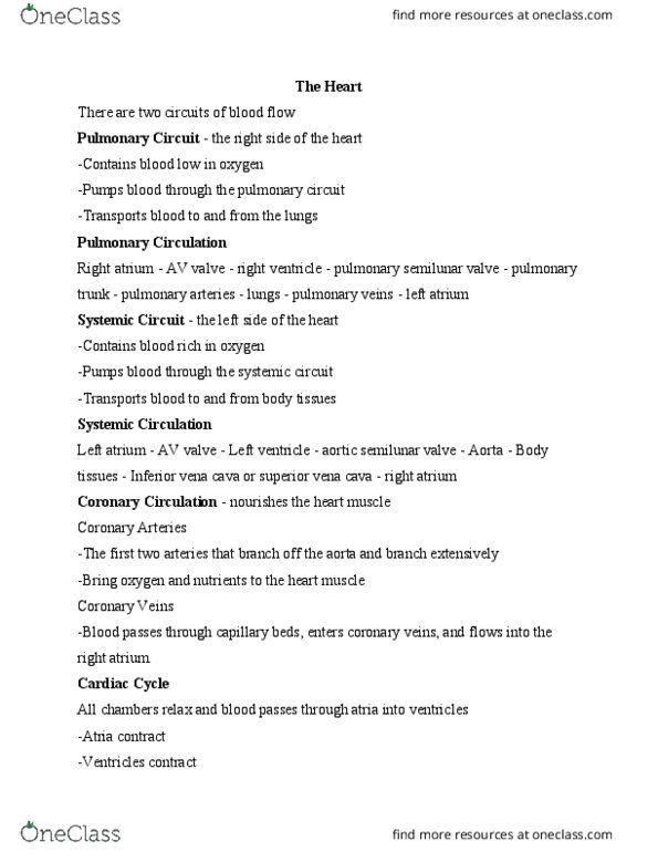
Chambers of the Heart
Atria: receiving chambers
-Small, thin-walled
-Convey blood to the ventricles
-Deoxygenated systemic blood goes to the right atrium via:
•Superior vena cava: brings systemic blood from above the diaphragm
•Inferior vena cava: brings systemic blood from below the diaphragm
•Coronary sinus: brings blood from the myocardium
Ventricles: discharging chambers
2

January 23, 2018
-These are the actual pumps of the heart
-Right ventricle: pumps blood to pulmonary trunk
-Left ventricle: pumps blood to the aorta
NB: won’t be given diagrams to label, but you need to know what the vessels do
Heart Valves
Blood flow is unidirectional - enforced by 4 heart valves
Atrioventricular (AV) valves: in between the atria and the ventricles
-Open downwards to allow blood to flow from the atria into the ventricles
-Prevent blood from flowing back into the atria when the ventricles start to contract
-Anchored by the chordae tendinae (attached to papillary muscles) to prevent them from opening upwards
(everting) when the ventricles contract
•Papillary muscles contract to pull on the chordae tedinae to close the valves during ventricular contraction
-Tricuspid valve: right AV valve
-Bicuspid valve: left AV valve
Semilunar valves: found in the major arteries leaving the heart
-Open upwards to allow blood to flow up out of the ventricles
-Prevent blood from flowing back into the ventricles after they have contracted
-Pulmonary valve
-Aortic valve
Valves open due to pressure differentials
-AV valves: open when the pressure is greater in the atria than in the ventricles
-Semilunar valves: open when the pressure is greater in the ventricles than in the vessels
Blood Pathway
Arrives into the right atrium
Flows into the right ventricle
Pumped out into the pulmonary trunk (splits into the right and left pulmonary arteries to the lungs)
After going to the lungs, it returns to left atrium via the pulmonary veins (4 in total - 2 per side)
Goes into the left ventricle
Pumped out of the heart, through the aorta, to the rest of the body
3
Document Summary
Role: bring oxygen and nutrients to tissues, remove wastes. Heart is simply 2 pumps, hollow blood vessels are delivery tubes. Arteries: carry blood away from the heart. Each beat of the heart (cardiac cycle) requires the coordination of many events. Pulmonary circulation: brings blood from the heart to the lungs for gas exchange: has serial blood ow - sequential changes to all of the blood following the pathway (ex. Systemic circulation: distributes oxygen-rich blood to the body, returns oxygen-deprived blood to the heart: has parallel blood ow - blood subdivided to service many vascular beds at one, relative distributions can be adjusted as per body"s need. Equal volumes owing in pulmonary and systemic circuits, but the two ventricles have unequal workloads. Pulmonary circuit (right ventricle): short, low pressure circulation: distance is short, so there is little resistance. Apex points to the left, oblique position in thorax ( left of midsternum) Pericardium: double-walled, broserous: fibrous pericardium: tough, outermost layer of ct.