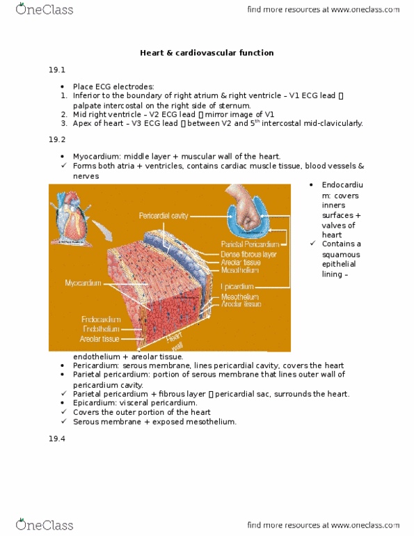
1) The external layer of the heart wall is the
Multiple Choice
endocardium.
myocardium.
epicardium.
parietal pericardium.
2) Check all that are characteristics of cardiac muscle.
Check All That Apply
Cells are long and cylindrical.
Cells are short and branching.
Cells have multiple nuclei at the periphery of the cell.
Cells have one or two nuclei in the center of the cell.
They are composed of thick and thin filaments.
The functional contractile unit is the sarcomere.
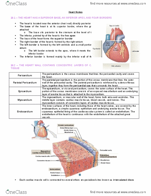
3) Match the chamber of the heart with the structure(s) from which it receives blood.
Coronary sinus
Pulmonary veins
Right atrium
Left atrium
Match each of the options above to the items below.
Right atrium
Right atrium Open choices for matching
Right ventricle
Right ventricle Open choices for matching
Left atrium
Left atrium Open choices for matching
Left ventricle
4) The less extensive distribution of T-tubules contributes to the more delayed onset and prolonged contraction of cardiac muscle tissue compared to skeletal muscle tissue.
True or False
5) The cells of the ___________ act as the heart's pacemaker, which establishes the pace for cardiac activity.
Multiple Choice
atrioventricular (AV) node
sinoatrial (SA) node
atrioventricular (AV) bundle
Purkinje cells
6) Match the cardiac vein with its description.
Travels close to the right marginal artery
In posterior interventricular sulcus
In anterior interventricular sulcus
In the posterior portion of cornary sulcus
Match each of the options above to the items below.
Great cardiac vein
Great cardiac vein Open choices for matching
Middle cardiac vein
Middle cardiac vein Open choices for matching
Small cardiac vein
Small cardiac vein Open choices for matching
Coronary sinus
7) Parasympathetic innervation to the heart comes from the medulla oblongata via the left and right ___________ nerves.
8) Check all that are components of the sympathetic innervation of the heart.
Check All That Apply
Cervical sympathetic ganglionCervical sympathetic ganglion
Spinal cordSpinal cord
Vagus nerve (CN X)Vagus nerve (CN X)
Cardiac nerveCardiac nerve
Sympathetic postganglionic axon
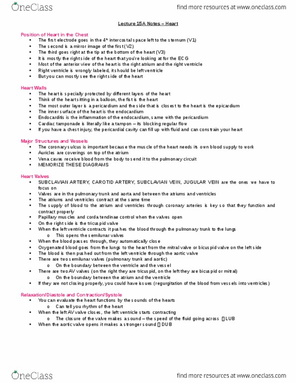
9) Check all that occur during ventricular contraction.
Check All That Apply
The AV valves open to allow blood to enter the ventricles from the atria.The AV valves open to allow blood to enter the ventricles from the atria.
The semilunar valves remain closed to prevent backflow of blood into the ventricles.The semilunar valves remain closed to prevent backflow of blood into the ventricles.
The semilunar valves open to allow blood to flow into the large arteries.The semilunar valves open to allow blood to flow into the large arteries.
The AV valves close to prevent backflow of blood into the atria.