BIO210Y5 Lecture Notes - Lecture 15: Coronary Circulation, Pulmonary Circulation, Intercostal Space

Document Summary
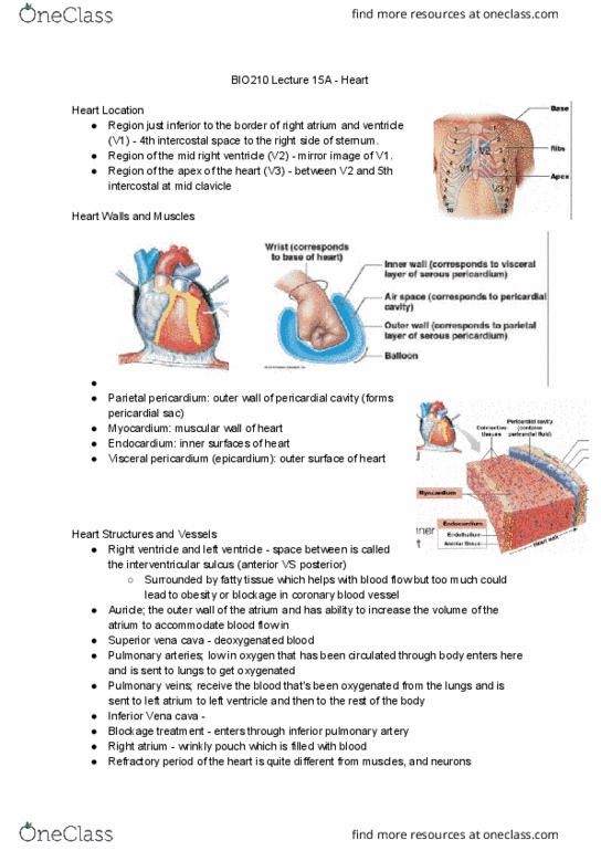
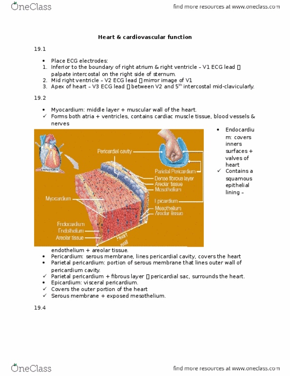
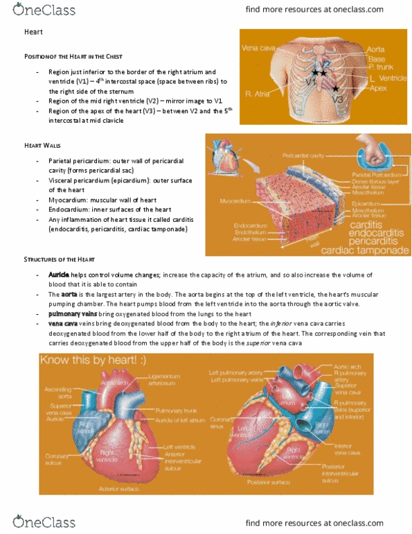
The first electrode goes in the 4th intercostal space left to the sternum (v1) The second is a mirror image of the first (v2) The third goes right at the tip at the bottom of the heart (v3) It is mostly the right side of the heart that you"re looking at for the ecg. Most of the anterior view of the heart is the right atrium and the right ventricle. Right ventricle is wrongly labeled, it should be left ventricle. But you can mostly see the right side of the heart. The heart is specially protected by different layers of the heart. Think of the heart sitting in a balloon, the fist is the heart. The most outer layer is a pericardium and the side that is closest to the heart is the epicardium. The inner surface of the heart is the endocardium. Endocarditis is the inflammation of the endocardium, same with the pericardium.