BIOB10H3 Lecture Notes - Lecture 9: Two-Dimensional Gel Electrophoresis, Celera Corporation, Polyacrylamide Gel Electrophoresis

14 Jun 2018
School
Department
Course
Professor

June 7, 2018
Lecture 9
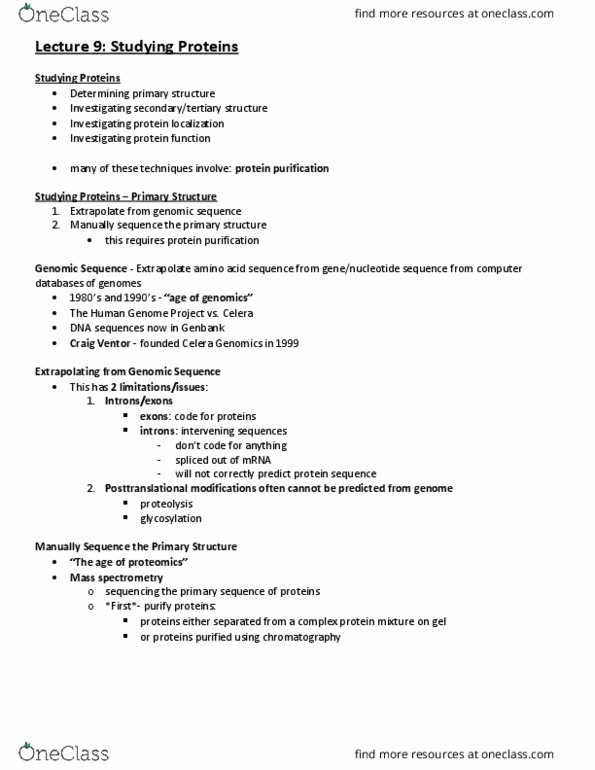
Studying proteins – Determining primary structure
• Extrapolate from genomic sequence (Database)
o Genomic sequence
▪ Extrapolate amino acid sequence from gene/nucleotide sequence from
computer database of genomes
▪ 1980’s and 1990’s- “age of genomics”
▪ The Human Genome Project (3.5B base pairs ~ 30K genes ~ each cell
makes 10K proteins) vs. Celera:
▪ Craig Ventor – founded Celera Genomics (company) in 1999
o Has 2 limitations/issues:
▪ Introns/exons
• exons: code for proteins
• introns: “intervening sequences”
o they don’t code for anything;
spliced out of mRNA; will not
correctly predict protein
sequence
• you read a gene, when you
translate it, you name an incorrect
protein because there’re introns
▪ Posttranslational modifications
often cannot be predicted from genome
• Proteolysis
• Glycosylation – adding sugars to protein
• Manually sequence the primary structure (you actually have it) – this requires
protein purification to get out the protein from the cell.
o “The age of proteomics” = we’re in this age now
o Mass spectrometry: sequencing the primary sequence of proteins;
o purify proteins either:
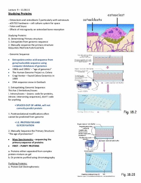
1. proteins either separated from a complex protein mixture on gel: Protein
Gel Electrophoresis: In 1D gel electrophoresis:
• cell lysates (break open cell) are made from cells of interest
• proteins are denatured by heat and put in tracking dye containing SDS.
SDS is (-) charged; it coats proteins and makes all proteins negatively
charged, so electrophoresis will pull the -ve protein
• proteins are separated on a gel based on size
find more resources at oneclass.com
find more resources at oneclass.com

• the gel is called polyacrylamide gel,
a porous gel (with many holes so it
moves on size)
• smaller proteins (low kDa) will move
through quickly and larger proteins
(high kDa) will move through very
slowly
• proteins move through by
electrophoresis: an electrical current
is applied through the gel – so there
is a positively charged end (anode)
and a negatively charged end
(cathode)
• proteins are all negative, so they
move towards the anode
• For 2D gel electrophoresis
(separation based on charge):
proteins are first separated
(Isoelectric focusing)
o Based on the charge of the proteins
• 1000’s of proteins can be resolved (separated) using 2D gel
electrophoresis
2. or proteins purified using
chromatography
• to isolate proteins from cell lysates
• lysates poured into column packed
with beads
• 3 major kinds of chromatography:
o gel-filtration chromatography -
> figure
▪ separates proteins based on
size; similar concept to gel electrophoresis
▪ big ones will move longer, and
small ones will come seconds
because large ones can’t go
through beads, but small ones get
inserted in the beads, so they
take longer to get separated – a
difference between gel electro
o ion-exchange chromatography -> figure
▪ separates proteins based on charge
find more resources at oneclass.com
find more resources at oneclass.com