BIO130H1 Lecture Notes - Lecture 4: Fluorescence Microscope, Intermediate Filament, Spindle Apparatus


31
BIO130H1 Full Course Notes
Verified Note
31 documents
Document Summary
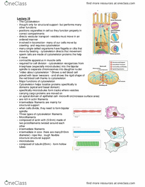
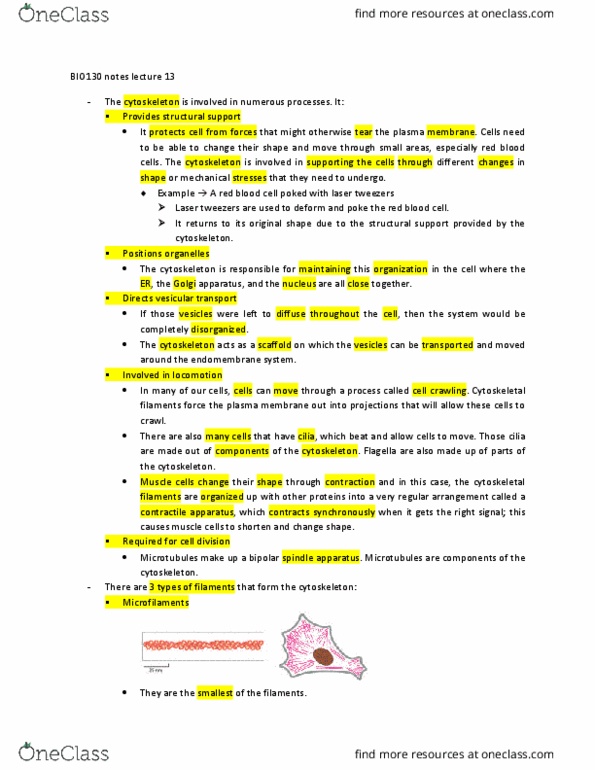
Reading: karp 7th edition, pages 324 345; 354 364. Required for cell division crawling along blood vessels, beating of cilia, flagella (both made up of cytoskeletal proteins), contractile motion of our mus cytoskeleton reorganizes e. mitotic spindle to separate chromosomes. A red blood cell poked with laser tweezers returns to its original shape due to the structural support provided by the cytoskeleton. Very often inside the cell along the periphery. A technique used to determine the location of specific proteins within the cell. An antibody is used which binds specifically to the protein of interest. A second antibody binds to the first antibody and is covalently tagged with a fluorescent molecule. A fluorescence microscope is used to excite the fluorescent molecule and visualise the light emitted. Note: you can also use primary antibody with fluorescent molecule but its more expensive. The light microscope has a resolution limit.