CSB351Y1 Lecture Notes - Lecture 20: Internal Ribosome Entry Site, Picornavirus, Vpg

26 May 2018
School
Department
Course
Professor

Lecture 20: Molecular Biology of Poliovirus
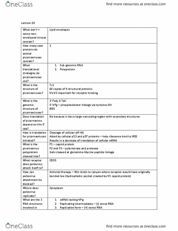
• Positive-sense non-enveloped viruses
• All have positive sense genomic RNA, acts directly as mRNA for protein synthesis and make protein
• Contains RNA, protein but no lipid envelopes
• Plant and bacterial viruses – single type of coat protein
• Animal picornaviruses contain four types of coat protein (V1, V2, V3, V4)
Phylogenetic Tree of Enteroviruses
• Rhino, Polio, Coxsackie, Entero, Aptho, Cardio, Hepato come from primodal virus (evolved from single ancestor)
Structure of Picornaviruses
• Icosahedral, T=1
• 27-30nm in diameter (very small), 4 structural proteins, 60 copies each per virion
• V1-3 form canyon where receptor binds), V4 inside where it contacts genomic RNA
Genome
• Ss + sense RNA, 7000-9000 nt in length (not that ig, Poly A tail at 3’ed
• VPg virio protei geoi ovaletly liked to 5’ ed i plae of ap
• Structural proteins (encoding for coat proteins) and non structure proteins area (proteases, replicase, etc)
• Contains IRES (internal ribosome entry site) – 8 AUG codons
Translation is cap-independent
• Pioravirus stops/ihiits ellular RNA’s traslatio through ap idig protei
- Cap binding protein inactivates mRNA translation by cleaving e1F-4G
- Translation of viral RNA can not proceed (decrease host mRNA translation)
• Poliovirus has IRES – internally initiates translation (P52 and P57 proteins bind IRES and allow ribosome binding)
Genomic Organization
• Monocistronic genome – contains one ORF encoding a large polyprotein
• 3C protease and 2A protease cleaves polyprotein → yields structural and non-structural proteins
Entry of picornaviruses to cells
• Picornaviruses inject RNA directly across plasma membrane after conformational change in capsid
• VP1 n-terminals pulled into the cell
• VPg with genomic RNA pulled into the cell, VP4 flips from above VP1 to underneath
Poliovirus proteins involved in replication
• Bind to CD155 receptor → ribosome bind to translate → polyprotein (+sense) → proteolytic cleavage → RdRp
to translate + to – sense RNA → multiple +sense RNA packaged with VP1-4 → leave cell
• All RNAs have VPg attahed to 5’ – VPg acts as primer for RNA synthesis (initiation)
• Cleaved proteins assemble into structure to become virus particle
• P1 → VP0, VP1, VP3 → 5S protomer → 14S pentamer → (80S procapsid + RNA) → 150S provirion → 150S virion
find more resources at oneclass.com
find more resources at oneclass.com
Document Summary
Phylogenetic tree of enteroviruses: rhino, polio, coxsackie, entero, aptho, cardio, hepato come from primodal virus (evolved from single ancestor) Icosahedral, t=1: 27-30nm in diameter (very small), 4 structural proteins, 60 copies each per virion, v1-3 form canyon where receptor binds), v4 inside where it contacts genomic rna. Translation is cap-independent: pi(cid:272)or(cid:374)avirus stops/i(cid:374)hi(cid:271)its (cid:272)ellular (cid:373)rna"s tra(cid:374)slatio(cid:374) through (cid:272)ap (cid:271)i(cid:374)di(cid:374)g protei(cid:374) Cap binding protein inactivates mrna translation by cleaving e1f-4g. Translation of viral rna can not proceed (decrease host mrna translation: poliovirus has ires internally initiates translation (p52 and p57 proteins bind ires and allow ribosome binding) Genomic organization: monocistronic genome contains one orf encoding a large polyprotein, 3c protease and 2a protease cleaves polyprotein yields structural and non-structural proteins. Entry of picornaviruses to cells: picornaviruses inject rna directly across plasma membrane after conformational change in capsid, vp1 n-terminals pulled into the cell, vpg with genomic rna pulled into the cell, vp4 flips from above vp1 to underneath.