CJH332H1 Lecture Notes - Lecture 10: Synaptic Vesicle, Snap25, Synaptobrevin


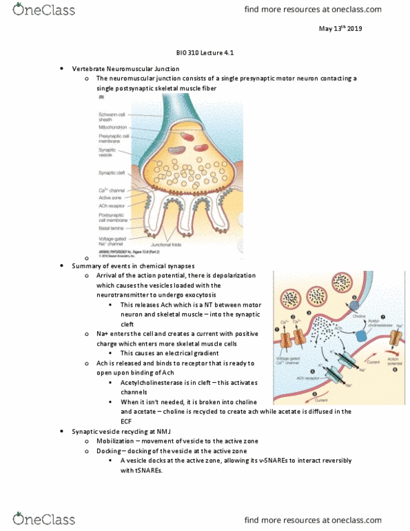
Lecture 10: Synaptic Activity and Synaptic Plasticity
• Both pre and pro synaptic areas have high density of proteins (not all t-SNAREs)
• Specific places vesicle fuses to
Exocytosis in more detail
• How does AP-induced Ca2+ entry actually cause vesicles to exocytose?
• Vesicles have proteins associated with them called v-SNAREs
• These interact with other proteins found on target presynaptic membrane (t-
SNAREs)
CAZ – dense and organize
• CAZ – cytomatrix of active zone
- Precise region closely localized to voltage gated Ca channels
- Specific sites of endocytosis (involving GTPases dynamin
pinching off presynaptic membrane)
- Some CAZ proteins remember presynaptic vesicle
- Vesicles reformed, filled with neurotransmitters, proteins
added back, etc
• Presynaptic element is very dense – multiple proteins linking each
other allowing docking at only specific places
Stepwise interactions of SNAREs
• Synaptic vesicle protein (v-SNARE) and target protein (t-SNARE)
• Presynaptic membrane protein SNAP-25 and syntaxin (HPC-1) – interacts with proteins on synaptic vesicle
• Munc18-1 prevents binding mode of syntaxin in the closed conformation
• Partial assembly of trans-SNARE complexes is facilitated by recently discovered chaperones
- Cysteine string proteins (CSPs) and synucleins enhance SNARE complex assembly
• Note that alpha-synuclein (enhancing formation of complexes) dysfunction is related to neurodegeneration
Stepwise interactions of SNAREs
• Vesicle associated SNAREs (v-SNAREs)
- Calcium sensor was deduced to be synaptotagmin (major) with synaptobrevin
- On synaptic vesicle membrane
• Synaptic membrane SNAREs (target t-SNAREs)
- SNAP-25 (folds back on itself) and syntaxin
- Tethers (include rab3a proteins) which include GTPases
- Munc18-1 holds things in a closed conformation (docked position)
- Primed position offers a 4 helix conformation (high affinity for each other)
◼ Syntaxin/synaptobrevin/2 arms of SNAP-25
- Complexin acts to stabilize this 4 helix structure util it’s ready to go
- AP → depolarization → VGCa open → Calcium binds to synaptotagmin
- Activates synaptotagmin to bind to the complex
- Complexin is displaced and pore forms/fusion of membranes
find more resources at oneclass.com
find more resources at oneclass.com
Document Summary
Lecture 10: synaptic activity and synaptic plasticity: both pre and pro synaptic areas have high density of proteins (not all t-snares, specific places vesicle fuses to. Exocytosis in more detail: how does ap-induced ca2+ entry actually cause vesicles to exocytose, vesicles have proteins associated with them called v-snares, these interact with other proteins found on target presynaptic membrane (t- Caz dense and organize: caz cytomatrix of active zone. Precise region closely localized to voltage gated ca channels. Specific sites of endocytosis (involving gtpases dynamin pinching off presynaptic membrane) Vesicles reformed, filled with neurotransmitters, proteins added back, etc: presynaptic element is very dense multiple proteins linking each other allowing docking at only specific places. Cysteine string proteins (csps) and synucleins enhance snare complex assembly: note that alpha-synuclein (enhancing formation of complexes) dysfunction is related to neurodegeneration. Stepwise interactions of snares: vesicle associated snares (v-snares) Calcium sensor was deduced to be synaptotagmin (major) with synaptobrevin.