Anatomy and Cell Biology 2221 Lecture Notes - Lecture 3: Dense Irregular Connective Tissue, Occipitalis Muscle, Frontalis Muscle

9 Dec 2016
School
Department
Professor
Document Summary
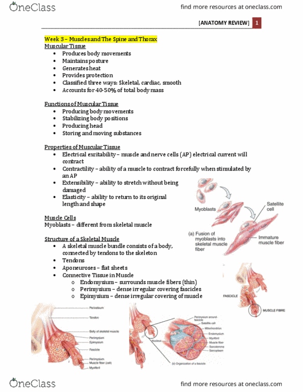
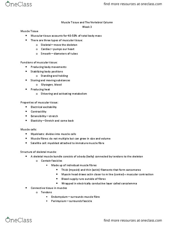
Muscular tissue accounts for 40 to 50% of total body mass. There are three types of muscular tissue: skeletal, cardiac, smooth. Myoblasts each skeletal muscle fiber arises from fusion of a hundred or more small mesodermal cells during embryonic development. Hyperplasia increasing total number of muscle fibers, only happens in fetal stage. Hypertrophy grow in size dramatically after birth. A skeletal muscle bundle consists of a body (belly) connected by tendons to the skeleton. Connective tissue in muscle: aponeuroses tendons arranged in flat sheets, endomysium thin wrapping of reticular fibers, perimysium dense irregular connective tissue covering fascicle, epimysium thicker covering of dense irregular connective tissue around periphery. Each muscle fiber has individual fibrous components, thick and thin changes that make up moving part of muscle (sarcomere) Each fascicle is bundle of individual cells. M line two myofilaments attach at middle of sarcomere. Contractile actin/myosin, generate force during contractions. Regulatory help switch muscle contraction process on and off.