Anatomy and Cell Biology 2221 Lecture Notes - Lecture 6: Medial Collateral Ligament, Rectus Femoris Muscle, Linea Aspera

9 Dec 2016
School
Department
Professor
Document Summary
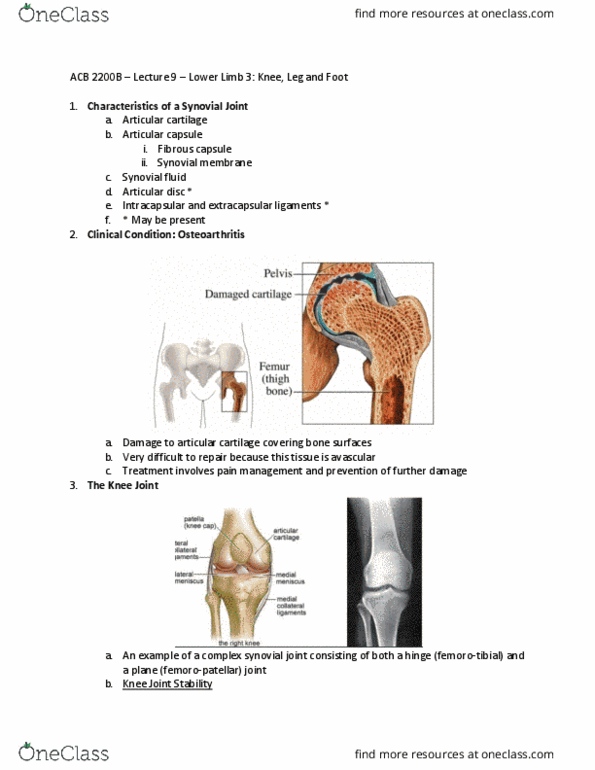
Patella is gateway from thigh to lower leg. Articulates with condyles of femur (hyland cartilage) Articular capsule: incomplete; thin sheath covering articulating bones. Extracapsular ligaments intracapsular ligaments: anterior and posterior cruciate. Articular discs (menisci: medial and lateral menisci. Bursae: prepatellar, infrapatellar and suprapatellar bursae. The anterior and posterior cruciate ligaments (acl/pcl) are intracapsular ligaments that stabilize the knee. Connect tibia to femur, cross and attach to opposite sides of condyle on femur. Acl prevents anterior sliding of tibia with respect to femur. Pcl prevents anterior sliding of femur with respect to tibia. Medial/lateral meniscus (shock absorption, prevent condyle from driving down into plate of tibia, increase surface area) Patellar ligament: continuation of common tendonous insertion of quadriceps femoris; strengthens anterior of joint. Oblique popliteal ligament: strengthens posterior surface of joint, hold synovial fluid into knee capsule. Tibial collateral ligament: strengthens medial aspect of joint; firmly attached to medial meniscus.