Anatomy and Cell Biology 2221 Lecture Notes - Lecture 8: Thoracic Duct, Suspensory Ligament, Referred Pain

31 Jan 2017
School
Department
Professor
Document Summary
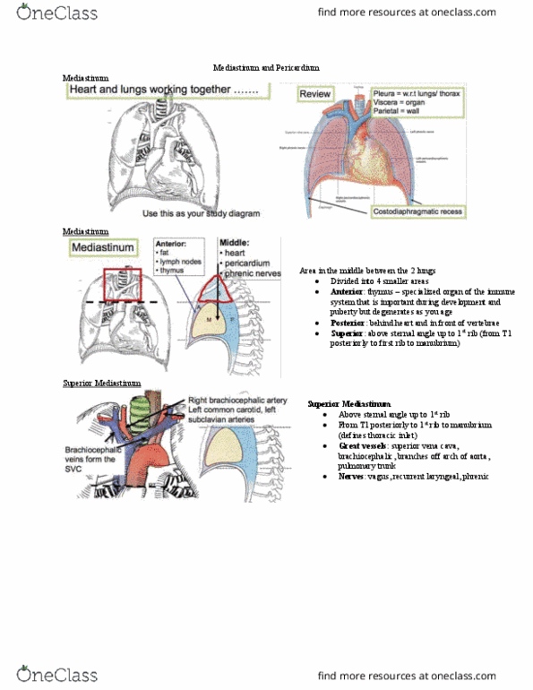
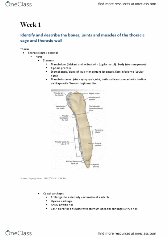
In many people, it stays as cartilage and never turns into bone. Costal cartilage & costal margin: costal margin- lower aspect of the rib cage (cartilage) T1 to margins of rib 1 and manubrium. Thoracic cage is the space in between these two outlets: defines the thorax for us. The thoracic cavity is the small space between the lining inside of the ribs and the lungs. Question: neck, shoulder, and arm pain, numbness and impaired circulation to the extremities (causing discoloration). The brachial plexus that is traveling through the axilla and the superior aperture and gets impingement. This known as the thoracic outlet syndrome. Divided into a mid-region of the thorax, called the mediastinum: main thing in here is the heart. Mediastinum: midline portion extends from sternum to vertebrae, and from superior to interior aperture. Superior aperture is defined by the first ribs. Heart is sitting in the mediastinum region.