Anatomy and Cell Biology 3309 Lecture Notes - Lecture 22: Lymph Node, Reticular Cell, Germinal Center

2 May 2018
School
Department
Professor

Histology Lecture
Lymph Nodes
November 30 2017
Question
- Which of the following is a way in which lymphatic vessels are different from veins?
o Only lymphatic vessels carry lymphocytes
o Lymphatic vessels have anchoring filaments
o Lymphatic vessels have valves
o Lymphatic vessels only move fluid in one direction
- All other ways, the lymphatic vessels and the veins are the same
Why are lymph nodes a prime site for cancer metastasis?
- Cancer calls may metastasize there – first place they look for in a cancer clinic
Lymphatic Nodule
- Concentrations of lymphocytes in a meshwork of reticular cells
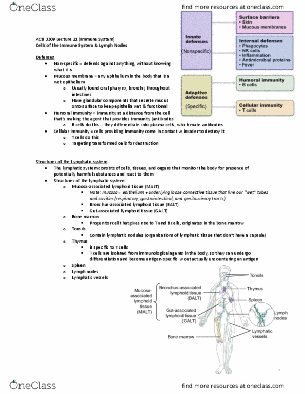
- Found in: GALT, BALT, lymph nodes, spleen and tonsils
- NOT encapsulated
- Has a very nice structure to it in comparison to the mucous associated lymphoid tissue (diffuse
tissue)
o This is nicely delineated
- LYMPHATIC NODULE ≠ LYMPH NODE
o Lymph node: small encapsulated lymph organs
o Lymphatic nodule: find throughout, collection of cells (lymphocytes) in organized fashion
▪ Mostly B-lymphocytes
- Have primary and secondary lymphatic nodule
- In image – it is a secondary lymphatic nodule:
o Has active germinal center with proliferating cells: middle of lymphatic nodule has lighter
staining part
▪ Lighter inside because cells actively undergoing proliferation: condensing nuclei,
dividing cellular organelles
find more resources at oneclass.com
find more resources at oneclass.com

o Cells on the outside are mostly B-lymphocytes = basophilic staining
- Lymphocytes are basically just nuclei when in circulation – nuclei take up most of the cell
o When they are proliferating, they are dividing their contents – do not have dark
concentration staining of nuclei from the lymphocytes
Primary vs. Secondary Lymphatic Nodules
- Primary:
o No germinal center, only mantle zones
o Lymphocytes not yet activated
o Few
- Secondary:
o Germinal AND mantle zones
▪ Germinal zone = lightly stained
• Cells are active; cytoplasm + eurchromatin of proliferating cells
▪ Mantle zone = darkly stained (more basophilic)
• B cell rich
o Lymphocytes activated (proliferation)
▪ Have encountered their antigen & undergoing proliferation
o Numerous
- These areas where we see lymphatic nodules, they are intercepting pathogens. Getting activated by
antigens & amount an immune response
o Vast majority of what we see are lymphatic nodules where the lymphocytes are activated &
proliferating
- WILL SEE MORE SECONDARY VS PRIMARY UNDER THE MICROSCOPE
- Tonsil = collection of lymph tissue
find more resources at oneclass.com
find more resources at oneclass.com

- Lymphatic nodule is different from the lymphoid diffuse tissue:
o Diffuse tissue – have few plasma cells, lymphocytes
here and there
o Lymphatic nodule – highly organized
Big picture summary
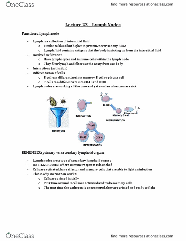
Functions of the Lymph Node
- Lymph = collection of fluid (interstitial fluid)
o Similar to blood BUT higher in protein
o NO RBC IN IT
o Contains all antigens the body is picking up from the interstitial fluid
- FUNCTIONS:
o Involved in filtration
▪ Filtering the lymph and pick up anything bad going throughout our body
o Interaction
o Activation
o Differentiation of cells
▪ B cell can differentiate into memory cell or plasma cells
▪ T cells also differentiate
- Lymph nodes are working ALL the time
- LYMPH NODES ARE SECONDARY LYMPHOID ORGANS
o Therefore, it is the battle ground – where you launch the immune response
find more resources at oneclass.com
find more resources at oneclass.com
Document Summary
Which of the following is a way in which lymphatic vessels are different from veins: only lymphatic vessels carry lymphocytes, lymphatic vessels have anchoring filaments, lymphatic vessels have valves, lymphatic vessels only move fluid in one direction. All other ways, the lymphatic vessels and the veins are the same. Cancer calls may metastasize there first place they look for in a cancer clinic. Concentrations of lymphocytes in a meshwork of reticular cells. Found in: galt, balt, lymph nodes, spleen and tonsils. In image it is a secondary lymphatic nodule: Lymphocytes are basically just nuclei when in circulation nuclei take up most of the cell: when they are proliferating, they are dividing their contents do not have dark concentration staining of nuclei from the lymphocytes. Primary: no germinal center, only mantle zones, lymphocytes not yet activated, few. These areas where we see lymphatic nodules, they are intercepting pathogens.