Anatomy and Cell Biology 3309 Lecture Notes - Lecture 26: Bronchiole, Hyaline Cartilage, Respiratory Epithelium

2 May 2018
School
Department
Professor

Histology Lecture 11 – Semester 2
Lungs
Learning Outcomes
- Identify the histological components of terminal bronchioles, respiratory bronchioles, and alveoli on
histological slides
- Define the unique histological features of terminal bronchioles, respiratory bronchioles, and alveoli
- List and describe the function of the cells making up terminal bronchioles, respiratory bronchioles,
and alveoli.
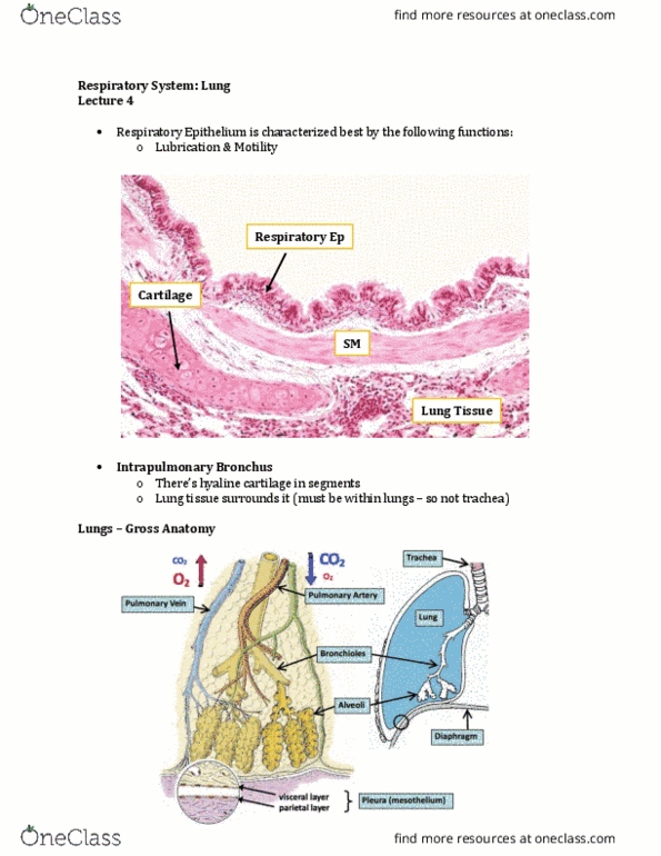
Review of Respiratory Passages
- Starting off with respiratory epithelium in the trachea, and as we move down the bronchi (moving
distally), we get simple columnar
- Further along you move from trachea through to intra-pulmonary bronchi, the less cartilage you
have involved
- Trachea – we have the trachealis muscle that helps control the diameter of the trachea during
inhalation and exhalation + allows food to go down the esophagus
- Lose the submucosa glands and BALT as we move down the respiratory tract
Epithelium
Cartilage
Smooth Muscle
Other
Trachea
Respiratory
Epithelium
Present in C shaped
rings (hyaline
cartilage)
Trachealis muscle
Contains
submucosa with
mucus secreting
glands
BALT
Bronchi
(Intra-
pulmonary)
Respiratory
EpitheliumSimple
Columnar
Discontinuous
cartilage plates
(hyaline cartilage)
Layer between lamina
propia and submucosa
(all the way around)
Contains
submucosa with
mucus secreting
glands
BALT
find more resources at oneclass.com
find more resources at oneclass.com

Respiratory Tract
- Two different types of bronchioles – terminal and respiratory
The Lung
- Trachea splits into extra-pulmonary bronchi (outside lungs) which then enter the lung, which are
now intra-pulmonary bronchi (inside lungs)
- Breathe in: rib cage comes out, diaphragm goes down
o Makes space inside the rib cage larger
o Lungs are adhered to the rib cage via a pleura – they follow the rib cage (fill up with air)
- Breathe out: space gets smaller and the air comes out
- Diaphragm muscle below the parietal layer + lung tissue above the visceral layer
- Two narrow line of cells (single layer of squamous cells) – epithelium
o Make up with pleura
o MESOTHELIUM
o Visceral layer + parietal layer (continuous with one another)
- Shiny layer on the lung = visceral layer of pleura
- Adhered to the diaphragm muscle and inside of rib cage = parietal layer
- There are vessels in the lung
o Point of us breathing is to oxygenate our blood
o Pulmonary artery – carries de-oxygenated blood
▪ ONLY ARTERY IN ADULT BODY THAT CRRIES DE-OXYGENATED BLOOD – coming
from the heart into the lung, picking up oxygen
o Pulmonary vein – carries oxygenated blood
▪ From the lung, to the heart to then get pumped to the rest of the body
- Bronchioles (start as intra-pulmonary bronchus)
o Terminal + respiratory bronchioles
o End with alveoli (end of the road for the air)
find more resources at oneclass.com
find more resources at oneclass.com

- Pulmonary artery and vein is coming to make a network of capillaries around the alveoli
Terminal Bronchioles
- When we talk about bronchioles instead of bronchi, we lose a few things
o General bronchioles: smaller airways (less than a mm)
▪ Lose cartilage and mucous glands
▪ Bronchioles no longer secrete mucous
▪ As you go down further your bronchioles, you get an increase in smooth muscle and
elastin
- First bronchioles after intra-pulmonary bronchus is the terminal bronchioles (red box)
- No cartilage (all bronchioles have no cartilage)
- Lots of smooth muscle
o Bands of smooth muscle running circumferentially around the bronchiole
- Elastin fibers running up and down (do not run perfectly parallel) - present in alveoli too
- Simple cuboidal epithelium
- Clara cells present
- Simple cuboidal epithelium with ciliated cell and Clara cells
o Clara cells secrete surfactant like lipoproteins preventing luminal adhesion in case airways
collapse during expiration
- Connective tissue with circumferential smooth muscle layer
find more resources at oneclass.com
find more resources at oneclass.com
Document Summary
Identify the histological components of terminal bronchioles, respiratory bronchioles, and alveoli on histological slides. Define the unique histological features of terminal bronchioles, respiratory bronchioles, and alveoli. List and describe the function of the cells making up terminal bronchioles, respiratory bronchioles, and alveoli. Starting off with respiratory epithelium in the trachea, and as we move down the bronchi (moving distally), we get simple columnar. Further along you move from trachea through to intra-pulmonary bronchi, the less cartilage you have involved. Trachea we have the trachealis muscle that helps control the diameter of the trachea during inhalation and exhalation + allows food to go down the esophagus. Lose the submucosa glands and balt as we move down the respiratory tract. Layer between lamina propia and submucosa (all the way around) Two different types of bronchioles terminal and respiratory. Trachea splits into extra-pulmonary bronchi (outside lungs) which then enter the lung, which are now intra-pulmonary bronchi (inside lungs)