Anatomy and Cell Biology 3309 Lecture Notes - Lecture 18: Suspensory Ligament, Ovarian Ligament, Fallopian Tube

15 May 2018
School
Department
Professor

Lecture 18 – Ovaries
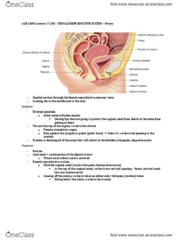
Female reproductive anatomy
- External genitalia: protect vaginal canal from debris or bacteria
o Labia minora
o Labia majora
- Clitoris: female stimulatory organ that rests against the symphysis pubis (pubic bone)
- Urethra: drains urine that collects in the bladder
- Rectum and anal canal: continuation of the digestive tract
- Female reproductive system:
o Vaginal canal receives penis during intercourse
o Cervical opening: forms cervical canal opening into uterine cavity (uterus)
- Fallopian tubes are on either side of the uterus
- Below the fallopian tubes are the ovaries
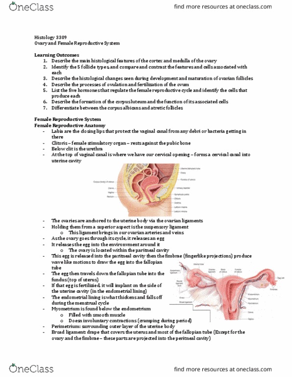
- Ovaries are anchored to the uterine body via ovarian ligament
- Holding them from a superior aspect is our suspensory ligament
- Suspensory ligament anchors ovary upwards which brings in ovarian arteries and veins
- Ovaries release eggs but not directly into the fallopian tubes
o It releases eggs into the environment surrounding it
o Ovaries are located within the peritoneal activity
- Eggs are released into the peritoneal cavity
- Fimbrae (finger-like projections) produce wave motion to draw egg cell into the fallopian
tube
- Egg travels down fallopian tube towards the body of the uterus and it enters at the fundus
(top of the uterus)
- If egg becomes fertilized, it implants on the side of the uterine cavity (on endometrial lining)
- Endometrial lining thickens during menstrual cycle and falls off during the cycle
- Myometrium is under the endometrium
o Thick muscular portion of the uterus
o Filled with smooth muscle and does involuntary contractions
o Cramping during period is involuntary contractions of the myometrium
find more resources at oneclass.com
find more resources at oneclass.com

- Perimetrium: surrounding/outer layer of uterine body
- Broad ligament: covers the entire uterus and most of the fallopian tubes (except the ovaries
and fimbrae)
o Ovaries and fimbrae are projected into the peritoneal cavity (where egg is released)
o Fimbrae creates sweeping motion to move the egg
Ovarian general structure
- Medulla is the core structure of the ovary
o Where ovarian arteries and veins enter into
o Nerves, lymphatic vessels, loose CT
- Cortex is surrounding the medulla
o Where ovarian follicles go through stages of development
o Find all of the ovarian follicles in the cortex
- Located within the medulla and cortex is ovarian stroma (CT of the ovary)
- On top of the cortex is germinal epithelium (single layer)
- Tunica albuginea (similar to male reproductive system) is connective tissue that wraps
around the ovary
find more resources at oneclass.com
find more resources at oneclass.com

- In the medulla there are cross sections of arteries and veins (NOT FOLLICLES)
- Cortex has follicles at different stages of development (primordial primary, mature
secondary, etc.)
Ovarian cortex structure
- There are ovarian follicles embedded in the ovarian stroma (ovarian stromal cells)
- Ovarian follicles are separate structures
- Boundary of ovarian cortex:
o Germinal epithelium: simple cuboidal
o Tunica albugina: below the epithelium is dense irrregular CT
Types of follicles (3)
- 1. Primordial follicles
find more resources at oneclass.com
find more resources at oneclass.com
Document Summary
External genitalia: protect vaginal canal from debris or bacteria: labia minora, labia majora. Clitoris: female stimulatory organ that rests against the symphysis pubis (pubic bone) Urethra: drains urine that collects in the bladder. Rectum and anal canal: continuation of the digestive tract. Female reproductive system: vaginal canal receives penis during intercourse, cervical opening: forms cervical canal opening into uterine cavity (uterus) Fallopian tubes are on either side of the uterus. Below the fallopian tubes are the ovaries. Ovaries are anchored to the uterine body via ovarian ligament. Holding them from a superior aspect is our suspensory ligament. Ovaries release eggs but not directly into the fallopian tubes. It releases eggs into the environment surrounding it. Suspensory ligament anchors ovary upwards which brings in ovarian arteries and veins: ovaries are located within the peritoneal activity. Eggs are released into the peritoneal cavity. Fimbrae (finger-like projections) produce wave motion to draw egg cell into the fallopian tube.