Anatomy and Cell Biology 3309 Lecture Notes - Lecture 5: Smooth Muscle Tissue, Cardiac Muscle Cell, Nuclear Membrane

22 May 2018
School
Department
Professor

Histology 3309
Cardiac & Smooth Muscle
Learning Objectives
- Desribe the organization of cardiac muscle fibers
- Describe the ultrastructure of cardiomyocytes
- Describe the components of the intercalated discs and their function
- Describe the organization of smooth muscle fibers
- Describe the process of smooth muscle contraction
- Compare the 3 muscle types in terms of structure and mechanism of contraction
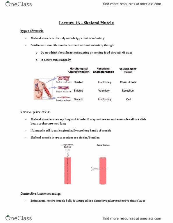
Review of skeletal muscle
- Multinucleated
- Nuclei located in the periphery (pushed to outside of sarcomeres)
- Striated – due to A and I band
- This is a cross section
- The arrow is pointing to the endomysium
o Loose connective tissue layer surround each muscle cell
- The muscle cells will create a fascicle, where we find a perimysium
- Then epimysium around the bundle of fascicles
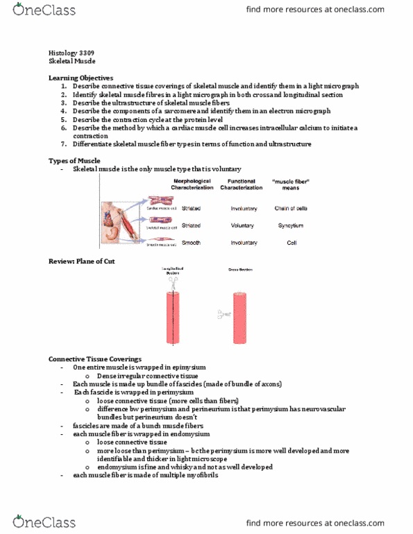
Type of Muscle
Type of Muscle
morphological
characterization
Functional
characterization
muscle fiber means
Cardiac (found in
myocardium layer of
heart)
Striated
Involuntary (controlled by
ANS)
Chain of muscle cells
Skeletal
Striated
Voluntary
Syncytium
Smooth (found in
hollow organs – ex: GI
tract, gallbladder,
urinary bladder, uterus)
Smooth
Involuntary (controlled by
ANS)
Cell
find more resources at oneclass.com
find more resources at oneclass.com

Cardia Muscle Fibres and Cardiomyocytes
- Rather than having one long cell with multiple nuclei, it has little tiny cells linked together to
create a muscle fiber
- Centrally located nuclei
- Mononucleated
o Sometimes, you might find a binucleated myocyte
- Surrounding the nucleus, we have the perinuclear cytoplasm (where we are going to find a lot of
the organelles)
o More specifically, organelles will be found at the poles of the cytoplasm (the ends)
o Aka perinuclear space
o In that space, there will be a lot mitochondria (bc this is such an energy dependent
process – heart is constantly beating)
- Muscle cells are going to be branched
o They can branch into or out of the screen
o They are still striated though
o We still have the actin-myosin overlap
- Depending on the section of cut, you may not see the perfect parallel striations
- Intercalated disks (the dark staining lines)
o Where the cardiac myocytes are linking together
o This is wehre you find fascia and macula adhera
o Very eosinophilic
o This is where our branched cardiac myocytes are linking together
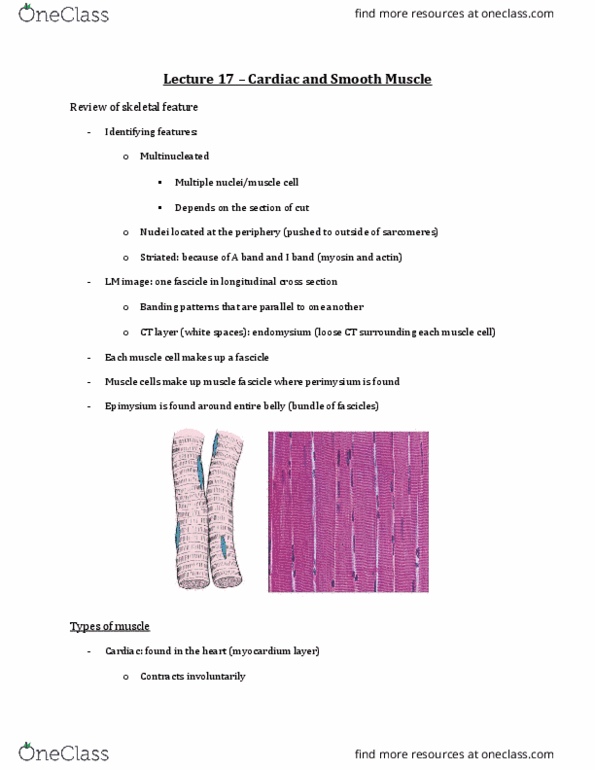
Plane of Cut?
- both of these are H&E
- right side is longitudinal section
o nucleus with perinuclear cytoplasm surrounding the nucleus
o can see striations (overlap bw actin and myosin)
o intercalated discs (as soon as you see these, you know its cardiac muscle)
- left side is cross section
o still have a centrally located nucleus surrounding by that actin and myosin
o can see some white space surrounding the nucleus
▪ so this is how we identify cardiac muscle in cross section – look for nuclei and
space around that nuclei
o may see stippling in cross section - its the myofibrils making up the cardiac myocyte
find more resources at oneclass.com
find more resources at oneclass.com

Cardiomyocyte Ultrastructure
- skeletal muscle
o peripherally located nucleus
o all the myofibrils making the myofiber
o sarcolemma and T tubules invaginating down and entering at the AI junction
o well developed SR (with specialized end of terminal cisternae creating that triad)
o T tubule is about same size as terminal cisternae
o SR is high developed and highly specialized
- cardiomyocyte
o branched pattern (kind of connected to eachother)
o centrally located nucleus
o T tubules of cardiomyocytes are much larger than T tubules of skeletal muscle
o T tubules are much larger than terminal cisternae
o T tubules enter at the Z line instead of the AI junction in the skeletal muscle
o SR is very sparse (not well developed)
▪ Looks like a few little strings hanging out
▪ This means that the terminal cisternae is also not very well developed
o SR and terminal cisternae create a DIAD
▪ Instead of a triad arrangement like the skeletal muscle
▪ Made of 1 small terminal cisternae and a large T tubule
Systemic Regulation of the Heart
- Cardiac muscle is myogenic (meaning it contracts on its own)
- Innervated by SYN(fight or flight) and PSYN (rest and digest)
- Mechanism of contraction is very similar to skeletal muscle - Overlap of actin and myosin
find more resources at oneclass.com
find more resources at oneclass.com